This page has been archived and is no longer updated
Discovery of DNA Structure and Function: Watson and Crick
Many people believe that American biologist James Watson and English physicist Francis Crick discovered DNA in the 1950s. In reality, this is not the case. Rather, DNA was first identified in the late 1860s by Swiss chemist Friedrich Miescher. Then, in the decades following Miescher's discovery, other scientists--notably, Phoebus Levene and Erwin Chargaff--carried out a series of research efforts that revealed additional details about the DNA molecule, including its primary chemical components and the ways in which they joined with one another. Without the scientific foundation provided by these pioneers, Watson and Crick may never have reached their groundbreaking conclusion of 1953: that the DNA molecule exists in the form of a three-dimensional double helix .

The First Piece of the Puzzle: Miescher Discovers DNA
Although few people realize it, 1869 was a landmark year in genetic research, because it was the year in which Swiss physiological chemist Friedrich Miescher first identified what he called "nuclein" inside the nuclei of human white blood cells. (The term "nuclein" was later changed to " nucleic acid " and eventually to " deoxyribonucleic acid ," or "DNA.") Miescher's plan was to isolate and characterize not the nuclein (which nobody at that time realized existed) but instead the protein components of leukocytes (white blood cells). Miescher thus made arrangements for a local surgical clinic to send him used, pus-coated patient bandages; once he received the bandages, he planned to wash them, filter out the leukocytes, and extract and identify the various proteins within the white blood cells. But when he came across a substance from the cell nuclei that had chemical properties unlike any protein, including a much higher phosphorous content and resistance to proteolysis (protein digestion), Miescher realized that he had discovered a new substance (Dahm, 2008). Sensing the importance of his findings, Miescher wrote, "It seems probable to me that a whole family of such slightly varying phosphorous-containing substances will appear, as a group of nucleins, equivalent to proteins" (Wolf, 2003).
More than 50 years passed before the significance of Miescher's discovery of nucleic acids was widely appreciated by the scientific community. For instance, in a 1971 essay on the history of nucleic acid research, Erwin Chargaff noted that in a 1961 historical account of nineteenth-century science, Charles Darwin was mentioned 31 times, Thomas Huxley 14 times, but Miescher not even once. This omission is all the more remarkable given that, as Chargaff also noted, Miescher's discovery of nucleic acids was unique among the discoveries of the four major cellular components (i.e., proteins, lipids, polysaccharides, and nucleic acids) in that it could be "dated precisely... [to] one man, one place, one date."
Laying the Groundwork: Levene Investigates the Structure of DNA
Meanwhile, even as Miescher's name fell into obscurity by the twentieth century, other scientists continued to investigate the chemical nature of the molecule formerly known as nuclein. One of these other scientists was Russian biochemist Phoebus Levene. A physician turned chemist, Levene was a prolific researcher, publishing more than 700 papers on the chemistry of biological molecules over the course of his career. Levene is credited with many firsts. For instance, he was the first to discover the order of the three major components of a single nucleotide (phosphate-sugar-base); the first to discover the carbohydrate component of RNA (ribose); the first to discover the carbohydrate component of DNA (deoxyribose); and the first to correctly identify the way RNA and DNA molecules are put together.
During the early years of Levene's career, neither Levene nor any other scientist of the time knew how the individual nucleotide components of DNA were arranged in space; discovery of the sugar-phosphate backbone of the DNA molecule was still years away. The large number of molecular groups made available for binding by each nucleotide component meant that there were numerous alternate ways that the components could combine. Several scientists put forth suggestions for how this might occur, but it was Levene's "polynucleotide" model that proved to be the correct one. Based upon years of work using hydrolysis to break down and analyze yeast nucleic acids, Levene proposed that nucleic acids were composed of a series of nucleotides, and that each nucleotide was in turn composed of just one of four nitrogen-containing bases, a sugar molecule, and a phosphate group. Levene made his initial proposal in 1919, discrediting other suggestions that had been put forth about the structure of nucleic acids. In Levene's own words, "New facts and new evidence may cause its alteration, but there is no doubt as to the polynucleotide structure of the yeast nucleic acid" (1919).
Indeed, many new facts and much new evidence soon emerged and caused alterations to Levene's proposal. One key discovery during this period involved the way in which nucleotides are ordered. Levene proposed what he called a tetranucleotide structure, in which the nucleotides were always linked in the same order (i.e., G-C-T-A-G-C-T-A and so on). However, scientists eventually realized that Levene's proposed tetranucleotide structure was overly simplistic and that the order of nucleotides along a stretch of DNA (or RNA) is, in fact, highly variable . Despite this realization, Levene's proposed polynucleotide structure was accurate in many regards. For example, we now know that DNA is in fact composed of a series of nucleotides and that each nucleotide has three components: a phosphate group ; either a ribose (in the case of RNA) or a deoxyribose (in the case of DNA) sugar; and a single nitrogen-containing base. We also know that there are two basic categories of nitrogenous bases: the purines ( adenine [A] and guanine [G]), each with two fused rings, and the pyrimidines ( cytosine [C], thymine [T], and uracil [U]), each with a single ring. Furthermore, it is now widely accepted that RNA contains only A, G, C, and U (no T), whereas DNA contains only A, G, C, and T (no U) (Figure 1).
Strengthening the Foundation: Chargaff Formulates His "Rules"
Erwin Chargaff was one of a handful of scientists who expanded on Levene's work by uncovering additional details of the structure of DNA, thus further paving the way for Watson and Crick. Chargaff, an Austrian biochemist, had read the famous 1944 paper by Oswald Avery and his colleague s at Rockefeller University, which demonstrated that hereditary units, or genes , are composed of DNA. This paper had a profound impact on Chargaff, inspiring him to launch a research program that revolved around the chemistry of nucleic acids. Of Avery's work, Chargaff (1971) wrote the following:
"This discovery, almost abruptly, appeared to foreshadow a chemistry of heredity and, moreover, made probable the nucleic acid character of the gene ... Avery gave us the first text of a new language, or rather he showed us where to look for it. I resolved to search for this text."
As his first step in this search, Chargaff set out to see whether there were any differences in DNA among different species . After developing a new paper chromatography method for separating and identifying small amounts of organic material, Chargaff reached two major conclusions (Chargaff, 1950). First, he noted that the nucleotide composition of DNA varies among species. In other words, the same nucleotides do not repeat in the same order, as proposed by Levene. Second, Chargaff concluded that almost all DNA--no matter what organism or tissue type it comes from--maintains certain properties, even as its composition varies. In particular, the amount of adenine (A) is usually similar to the amount of thymine (T), and the amount of guanine (G) usually approximates the amount of cytosine (C). In other words, the total amount of purines (A + G) and the total amount of pyrimidines (C + T) are usually nearly equal. (This second major conclusion is now known as "Chargaff's rule.") Chargaff's research was vital to the later work of Watson and Crick, but Chargaff himself could not imagine the explanation of these relationships--specifically, that A bound to T and C bound to G within the molecular structure of DNA (Figure 2).
Putting the Evidence Together: Watson and Crick Propose the Double Helix
Chargaff's realization that A = T and C = G, combined with some crucially important X-ray crystallography work by English researchers Rosalind Franklin and Maurice Wilkins, contributed to Watson and Crick's derivation of the three-dimensional, double-helical model for the structure of DNA. Watson and Crick's discovery was also made possible by recent advances in model building, or the assembly of possible three-dimensional structures based upon known molecular distances and bond angles, a technique advanced by American biochemist Linus Pauling. In fact, Watson and Crick were worried that they would be "scooped" by Pauling, who proposed a different model for the three-dimensional structure of DNA just months before they did. In the end, however, Pauling's prediction was incorrect.
Using cardboard cutouts representing the individual chemical components of the four bases and other nucleotide subunits, Watson and Crick shifted molecules around on their desktops, as though putting together a puzzle. They were misled for a while by an erroneous understanding of how the different elements in thymine and guanine (specifically, the carbon, nitrogen, hydrogen, and oxygen rings) were configured. Only upon the suggestion of American scientist Jerry Donohue did Watson decide to make new cardboard cutouts of the two bases, to see if perhaps a different atomic configuration would make a difference. It did. Not only did the complementary bases now fit together perfectly (i.e., A with T and C with G), with each pair held together by hydrogen bonds, but the structure also reflected Chargaff's rule (Figure 3).
Although scientists have made some minor changes to the Watson and Crick model, or have elaborated upon it, since its inception in 1953, the model's four major features remain the same yet today. These features are as follows:
- DNA is a double-stranded helix, with the two strands connected by hydrogen bonds. A bases are always paired with Ts, and Cs are always paired with Gs, which is consistent with and accounts for Chargaff's rule.
- Most DNA double helices are right-handed; that is, if you were to hold your right hand out, with your thumb pointed up and your fingers curled around your thumb, your thumb would represent the axis of the helix and your fingers would represent the sugar-phosphate backbone. Only one type of DNA, called Z-DNA , is left-handed.
- The DNA double helix is anti-parallel, which means that the 5' end of one strand is paired with the 3' end of its complementary strand (and vice versa). As shown in Figure 4, nucleotides are linked to each other by their phosphate groups, which bind the 3' end of one sugar to the 5' end of the next sugar.
- Not only are the DNA base pairs connected via hydrogen bonding, but the outer edges of the nitrogen-containing bases are exposed and available for potential hydrogen bonding as well. These hydrogen bonds provide easy access to the DNA for other molecules, including the proteins that play vital roles in the replication and expression of DNA (Figure 4).
One of the ways that scientists have elaborated on Watson and Crick's model is through the identification of three different conformations of the DNA double helix. In other words, the precise geometries and dimensions of the double helix can vary. The most common conformation in most living cells (which is the one depicted in most diagrams of the double helix, and the one proposed by Watson and Crick) is known as B-DNA . There are also two other conformations: A-DNA , a shorter and wider form that has been found in dehydrated samples of DNA and rarely under normal physiological circumstances; and Z-DNA, a left-handed conformation. Z-DNA is a transient form of DNA, only occasionally existing in response to certain types of biological activity (Figure 5). Z-DNA was first discovered in 1979, but its existence was largely ignored until recently. Scientists have since discovered that certain proteins bind very strongly to Z-DNA, suggesting that Z-DNA plays an important biological role in protection against viral disease (Rich & Zhang, 2003).
Watson and Crick were not the discoverers of DNA, but rather the first scientists to formulate an accurate description of this molecule's complex, double-helical structure. Moreover, Watson and Crick's work was directly dependent on the research of numerous scientists before them, including Friedrich Miescher, Phoebus Levene, and Erwin Chargaff. Thanks to researchers such as these, we now know a great deal about genetic structure, and we continue to make great strides in understanding the human genome and the importance of DNA to life and health.
References and Recommended Reading
Chargaff, E. Chemical specificity of nucleic acids and mechanism of their enzymatic degradation. Experientia 6 , 201–209 (1950)
---. Preface to a grammar of biology. Science 171 , 637–642 (1971)
Dahm, R. Discovering DNA: Friedrich Miescher and the early years of nucleic acid research. Human Genetics 122 , 565–581 (2008)
Levene, P. A. The structure of yeast nucleic acid. IV. Ammonia hydrolysis . Journal of Biological Chemistry 40 , 415–424 (1919)
Rich, A., &. Zhang, S. Z-DNA: The long road to biological function. Nature Reviews Genetics 4 , 566–572 (2003) ( link to article )
Watson, J. D., & Crick, F. H. C. A structure for deoxyribose nucleic acid. Nature 171 , 737–738 (1953) ( link to article )
Wolf, G. Friedrich Miescher: The man who discovered DNA. Chemical Heritage 21 , 10-11, 37–41 (2003)
- Add Content to Group
Article History
Flag inappropriate.


Email your Friend

- | Lead Editor: Bob Moss

Within this Subject (34)
- Applications in Biotechnology (4)
- Discovery of Genetic Material (4)
- DNA Replication (6)
- Gene Copies (5)
- Jumping Genes (4)
- RNA (7)
- Transcription & Translation (4)
Other Topic Rooms
- Gene Inheritance and Transmission
- Gene Expression and Regulation
- Nucleic Acid Structure and Function
- Chromosomes and Cytogenetics
- Evolutionary Genetics
- Population and Quantitative Genetics
- Genes and Disease
- Genetics and Society
- Cell Origins and Metabolism
- Proteins and Gene Expression
- Subcellular Compartments
- Cell Communication
- Cell Cycle and Cell Division
© 2014 Nature Education
- Press Room |
- Terms of Use |
- Privacy Notice |

Visual Browse
Watson and Crick DNA Model
History of DNA Double Helical Structure
4 key features of watson and cricks dna model, components of dna, the nitrogen bases or nucleotides, deoxyribose sugar, the phosphate group (phosphate backbone), physical properties of dna, chemical properties of dna, features of the watson-crick structure of dna, watson-crick model of dna summary, biological importance of dna, how dna strands are connected to each other, chargaff’s rule, what are the key features of the watson and crick dna model, how did chargaff’s rules contribute to the development of the watson and crick model, what was the significance of watson and crick’s discovery of the double helix structure, how does the watson and crick model explain the process of dna replication, what is the role of complementary base pairing in the watson and crick dna model, what evidence or data did watson and crick use to support their model of dna, how did the watson and crick model of dna revolutionize our understanding of genetics and molecular biology, here are 10 recent research findings on dna models, what model of dna was discovered by watson and crick, what did watson and crick’s model of dna show, which description best explains watson and crick’s dna model, how to draw watson and crick’s model of dna, how did chargaff’s rules help watson and crick’s model of dna, how did watson and crick develop the model of dna, what did watson and crick’s model of dna look like, what does watson and crick’s model of dna demonstrate, when did watson and crick present their dna model, why was watson and crick’s first dna model incorrect.
In 1953, James Watson and Francis Crick made a groundbreaking contribution to the understanding of DNA structure. By combining physical and chemical data, they proposed a model for DNA as a double helix, a twisted molecule consisting of two complementary strands held together by hydrogen bonds. This model revolutionized our knowledge of genetics and unlocked the genetic code that underlies all living organisms.
The Watson and Crick model revealed that DNA serves as the backbone for all life forms. It demonstrated that the structure of DNA contains genetic information and can make copies of itself, which are then passed down through generations. This model provided a key insight into how DNA functions and plays a fundamental role in the transmission of genetic traits.
The Watson and Crick model, also known as the double helix model, represented a scientific breakthrough of immense significance. It explained that DNA is composed of two strands, each consisting of a sugar-phosphate backbone and nitrogenous bases. These bases, including adenine, thymine, guanine, and cytosine, pair up through hydrogen bonds to form the rungs of the DNA ladder. This model not only explained the structure of DNA but also how it replicates and carries out essential biological processes.
Today, the Watson and Crick model remains a fundamental concept in molecular biology. It provides one of the best visual representations of the structure of double-helix DNA. DNA itself is a polymer composed of monomer units called deoxyribonucleotides, which are linked together by phosphodiester bonds. Many scientists, including Friedrich Miescher, P.A. Levene, W.T. Astbury, Maurice Wilkins, and Rosalind Franklin, contributed to the understanding of DNA’s components and composition, laying the groundwork for Watson and Crick’s breakthrough.
The significance of Watson and Crick’s model was widely recognized, leading to their receipt of the Nobel Prize in Physiology or Medicine in 1962. They shared this prestigious award with Maurice Wilkins, although Rosalind Franklin, who made significant contributions to the understanding of DNA structure, tragically passed away in 1958 and thus did not receive the recognition she deserved.
The history of the DNA double helical structure is a remarkable journey of scientific discovery that involved several key scientists and breakthroughs. Here is a timeline highlighting the significant events:
1869: Freidrich Miescher, a Swiss physiological chemist, identified a substance in the nuclei of white blood cells , which he named “Nuclein.” This discovery laid the foundation for the understanding of nucleic acids, such as deoxyribonucleic acid (DNA).
1920: Phoebus Aaron Theodore Levene, an American organic chemist, determined the elemental composition of nucleic acids. He proposed the “Tetranucleotide hypothesis,” suggesting that the base composition of all four nitrogenous bases (A, T, G, and C) would be the same.
1940: William Thomas Astbury, an English physicist and molecular biologist, utilized X-ray crystallography to present a three-dimensional model of DNA. This model provided insights into the structural characteristics of DNA.
1950: Erwin Chargaff, an Austro-Hungarian biochemist, made significant contributions to the understanding of DNA biochemistry. He formulated two key principles known as “Chargaff’s Rule.” The first principle stated that the sum of purines (A and G) is equal to the sum of pyrimidines (T and C) in DNA. The second principle highlighted variations in DNA composition between different species.
1952: Maurice Wilkins, Rosalind Franklin, and their colleagues utilized X-ray diffraction to obtain photographs of DNA. Rosalind Franklin’s super X-ray diffraction photograph provided crucial insights into the structure of DNA.
1953: James Watson and Francis Crick, working in collaboration with Wilkins and Franklin, unveiled the double-helical structure of DNA. Watson and Crick’s model, based on the X-ray diffraction data, provided a groundbreaking understanding of how DNA is structured. This discovery was widely acclaimed and earned them the Nobel Prize in Physiology or Medicine in 1962, which they shared with Wilkins.
The discovery of the DNA double helical structure revolutionized our understanding of genetics and paved the way for advancements in molecular biology. The contributions of these scientists laid the foundation for further research in genetics, DNA replication , and the decoding of the genetic code, making significant contributions to the field of biological sciences.
The Watson and Crick model of DNA, which revolutionized our understanding of genetics, is characterized by four major features:
- Complementary Base Pairing: The DNA molecule consists of two individual strands of polynucleotides that are connected by hydrogen bonds. The nucleotide bases in DNA, adenine (A), thymine (T), guanine (G), and cytosine (C), exhibit a complementary pairing rule: purine bases (A and G) always pair with pyrimidine bases (T and C). This base pairing allows for the precise replication and transmission of genetic information.
- Right-Handed Double Helix: In most cases, the double helix structure of DNA is right-handed, meaning it twists in a clockwise direction when viewed along its axis. It completes one full turn every 34 angstroms (34A°). Visualizing the double helix, if you hold your right thumb pointed up to represent the axis, the sugar-phosphate backbones run along the outside of the molecule, while the nitrogen bases are positioned inside. An example of a left-handed DNA structure is the Z-DNA, which is an exception to the right-handed helix.
- Anti-Parallel Strands: The two strands of DNA in the double helix are anti-parallel to each other. This means that if one strand has a starting point at the 5′ end, the other strand will have its starting point at the 3′ end, and vice versa. This arrangement is due to the polarity of the phosphodiester linkage , which connects the nucleotides . The anti-parallel nature of the strands is crucial for DNA replication and transcription processes.
- Hydrogen Bonding and Accessibility: The outer exposed molecules of the nitrogen bases in DNA have the potential to form hydrogen bonds with other molecules. These hydrogen bonds play a crucial role in DNA replication and gene expression . They provide easy access to the DNA molecule, allowing enzymes and proteins to interact with specific base sequences for processes such as DNA replication, transcription, and protein synthesis.
These four major features of the Watson and Crick DNA model—complementary base pairing, right-handed double helix, anti-parallel strands, and hydrogen bonding—provide a comprehensive understanding of the structure and function of DNA. They explain how genetic information is stored, replicated, and expressed, and have paved the way for significant advancements in the field of molecular biology.
DNA, or deoxyribonucleic acid, is composed of specific components that together form the structure of the molecule. These components include nucleotide monomers, which are the building blocks of DNA.
- The nucleotide monomers in DNA consist of three molecular parts: nitrogen bases, deoxyribose sugar, and a phosphate group. There are four types of nitrogen bases found in DNA: adenine (A), cytosine (C), thymine (T), and guanine (G). These bases are responsible for carrying the genetic information in the DNA molecule.
- The deoxyribose sugar molecule is a five-carbon sugar that forms the backbone of the DNA strand. Each nucleotide in the DNA chain is connected to the adjacent nucleotide through the deoxyribose sugar. The deoxyribose sugar provides structural stability to the DNA molecule.
- The phosphate group is attached to the deoxyribose sugar in each nucleotide. It consists of a phosphorus atom bonded to four oxygen atoms. The phosphate groups link together to form a phosphate backbone, running along the outer edges of the DNA strands. The phosphate backbone provides additional structural support and stability to the DNA molecule.
In summary, the components of DNA include nucleotide monomers, which consist of nitrogen bases (adenine, cytosine, thymine, and guanine), deoxyribose sugar, and a phosphate group. These components combine to form the unique structure of DNA and encode the genetic information that is essential for all living organisms.
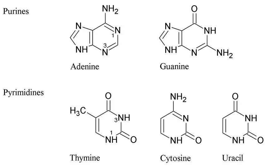
- The nitrogen bases, or nucleotides, play a crucial role in the structure and function of DNA. DNA strands are made up of these monomers, which are often referred to as bases due to their cyclic organic nature.
- There are four different nucleotides in DNA: adenine (A), thymine (T), cytosine (C), and guanine (G). These nucleotides join together to form a DNA strand, with the base portions projecting inward from the backbone of the strand.
- In the DNA double helix structure, two strands bind together via the nitrogen bases and twist around each other, forming a double helix. The nitrogen bases exhibit a specific pairing pattern.
- Adenine always pairs with thymine (A-T) via two hydrogen bonds, while cytosine always pairs with guanine (C-G) via three hydrogen bonds. This complementary base pairing ensures that the amount of adenine is equal to the amount of thymine, and the amount of guanine is equal to the amount of cytosine. The hydrogen bonding between the base pairs stabilizes the structure of the DNA double helix.
- The nitrogen bases in DNA are divided into two categories: purines and pyrimidines. Adenine and guanine are purines, while cytosine and thymine are pyrimidines. These bases form the foundation of the nucleotides and ultimately the DNA molecule.
- The shape of the DNA helix is stabilized by hydrogen bonding and hydrophobic interactions between the bases. The hydrogen atoms of the amino groups act as donors, while the carbonyl oxygen and ring nitrogen act as acceptors in the hydrogen bonding interactions.
- The DNA double helix structure contains ten nucleotides in each turn, with an internucleotide distance of 3.4 angstroms (3.4A°). The full turn of the helix has a length of 34 angstroms (34A°) and a diameter of approximately 20 angstroms (20A°).
- This helical structure gives rise to two grooves: the major groove and the minor groove. The major groove is deep and wide, serving as the specific binding site for proteins involved in DNA interactions. On the other hand, the minor groove is the narrower space between the two strands.
- In summary, the nitrogen bases or nucleotides in DNA are crucial components that contribute to the structure and function of the molecule. They form complementary base pairs, have specific hydrogen bonding patterns, and stabilize the DNA double helix.
- The nucleotide bases adenine, thymine, cytosine, and guanine are fundamental to the genetic code and play a vital role in DNA replication, transcription, and protein synthesis .
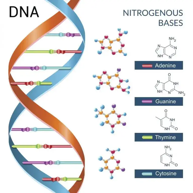
- Deoxyribose sugar, also known as 2-deoxyribose, is a pentose sugar that serves as a key component of deoxyribonucleic acid (DNA). It is derived from the pentose sugar ribose and has the chemical formula C5H10O4.
- In DNA, deoxyribose sugar forms the backbone of the molecule, alternating with phosphate groups. It binds to the nitrogenous bases adenine, thymine, guanine, and cytosine, which are responsible for carrying the genetic information in DNA. The absence of one hydroxyl group on the 2′ carbon atom distinguishes deoxyribose from ribose.
- Deoxyribose sugar plays a critical role in DNA, which represents the genetic information in all living cells. It is an essential component of life and is shared among all living organisms.
- The deoxyribose sugar molecule consists of five carbon atoms, with four of them forming carbon molecules and one oxygen molecule arranged cyclically. Its flexible structure allows it to adopt various conformations, such as the C2′ endo configuration observed in the canonical B-DNA.
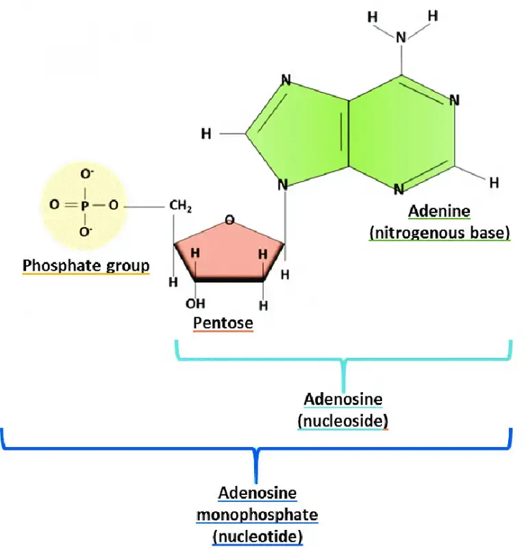
- When a nucleobase is attached to a deoxyribose sugar, it forms a nucleoside . The 1′ carbon of the pentose sugar bonds with the nitrogenous base, while the 5′ carbon atom bonds to the phosphate group. These bonds are facilitated by hydrogen bonding.
- During DNA replication, enzymes are involved in breaking the hydrogen bonds, resulting in the separation of the DNA strands. The newly formed deoxyribose molecules then attach to nitrogenous bases and phosphate groups, ultimately forming a new DNA molecule. The process of replication ensures the accurate transmission of genetic information from one generation to the next.
- In summary, deoxyribose sugar is a vital component of DNA. It forms the backbone of the DNA molecule, binding to nitrogenous bases and phosphate groups. Its unique structure and bonding patterns contribute to the stability and functionality of DNA, enabling the storage and transmission of genetic information.
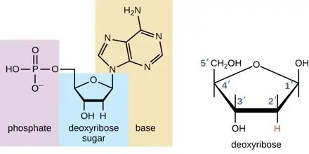
- The phosphate group, also known as the phosphate backbone, plays a critical role in the structure and function of nucleic acids, including DNA. It forms the structural framework of DNA, providing stability and defining the directionality of the molecule.
- The sugar-phosphate backbone is composed of alternating sugar and phosphate groups. In DNA, nucleotides are linked together in a chain by ester bonds between the sugar base of one nucleotide and the phosphate group of the adjacent nucleotide. The sugar is referred to as the 3′ end, while the phosphate is the 5′ end of each nucleotide.
- The phosphate group attached to the 5′ carbon of the sugar on one nucleotide forms an ester bond with the free hydroxyl group on the 3′ carbon of the next nucleotide. These bonds are known as phosphodiester bonds. As DNA is synthesized, the sugar-phosphate backbone extends or grows in the 5′ to 3′ direction.
- In double-stranded DNA, the sugar-phosphate backbones of the two strands run opposite each other, and they twist together to form the characteristic double-helix shape. This twisting and alignment of the sugar-phosphate backbones contribute to the stability and structural integrity of DNA.
- The phosphate group carries a negative charge, making the sugar-phosphate backbone negatively charged as well. This negative charge and the hydrophilic nature of the phosphate group allow the DNA backbone to interact with water molecules.
- The phosphate group is an essential component of every single strand of the DNA molecule. Its chemical structure consists of a phosphorus atom bonded with four oxygen atoms, with three single bonds and one double bond.
- Apart from its structural role, the phosphate group also functions as an energy donor during DNA synthesis. The release of phosphate groups provides energy for the formation of phosphodiester bonds between nucleotides.
- In summary, the phosphate group forms the backbone of DNA, linking nucleotides together through phosphodiester bonds. It provides stability, directionality, and the necessary negative charge for the molecule. The phosphate group plays a fundamental role in DNA structure, function, and the transmission of genetic information.
Watson and Crick model of DNA
Watson and Crick exhibited the structure of DNA after examining the manuscript of Linus Pauling and Corey. Linus Pauling and Corey presented the unsuccessful 3D structure of nucleic acid in 1953. Then, (in early 1953), Watson and Crick proposed a double-helical structure for DNA by combining physical and chemical property data. The main characteristics of the DNA model developed by Watson and Crick include:
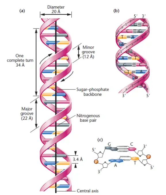
DNA, according to the Watson and Crick model, exhibits several physical properties that contribute to its structure and function:
- Double-stranded helix: DNA consists of two polynucleotide chains that are spirally twisted around each other, giving it a twisted ladder-like appearance. This double-stranded helical structure provides stability to the DNA molecule.
- Antiparallel polarity: The two polynucleotide strands of DNA run in opposite directions, resulting in antiparallel polarity. One strand runs in the 5′-3′ direction, while the other runs in the 3′-5′ direction. This arrangement allows for complementary base pairing between the strands.
- Diameter: The diameter of the DNA double helix is approximately 20Å (angstroms). This consistent diameter is maintained due to the specific pairing of nucleotide bases within the helix.
- Internucleotide distance: The distance between two adjacent nucleotides, known as the internucleotide distance, is approximately 3.4Å. This distance refers to the spacing between the individual base pairs along the DNA molecule.
- Length of helix and base pairs per turn: A complete turn of the DNA helix has a length of approximately 34Å and consists of 10 base pairs. This characteristic length and base pairing arrangement contribute to the overall stability and structure of DNA.
- Right-handed twist: DNA exhibits a right-handed twist or a clockwise direction when viewed from the top. This twist allows the two strands to wrap around each other in a helical manner.
- Major and minor grooves: The twisting of the DNA strands creates distinct features known as major and minor grooves. The major groove is wider, while the minor groove is narrower. These grooves provide binding sites for various proteins involved in DNA replication, transcription, and other cellular processes.
Understanding the physical properties of DNA, such as its helical structure, diameter, and grooves, is crucial for comprehending its role in genetic information storage, replication, and gene expression. These properties enable DNA to function as the fundamental molecule of heredity in living organisms.
DNA exhibits various chemical properties that are crucial to its structure and function:
- Nucleotide bases: DNA is composed of four nucleotide bases: adenine (A), guanine (G), cytosine (C), and thymine (T). Adenine and guanine are purine bases, characterized by a single-ring structure, while cytosine and thymine are pyrimidine bases, featuring a double-ring structure.
- Complementary base pairing: The two DNA strands are joined together through complementary base pairing of the nucleotide bases. Adenine pairs with thymine through two hydrogen bonds, while guanine pairs with cytosine through three hydrogen bonds. This specific base pairing forms the foundation of DNA’s double-stranded structure.
- Hydrogen bonding: The nucleotide bases within the DNA strands are held together by strong hydrogen bonds. These hydrogen bonds contribute to the stability and integrity of the DNA molecule.
- Chargaff’s rule: The base composition of DNA follows Chargaff’s rule, which states that the sum of purines (A and G) is equal to the sum of pyrimidines (C and T). Thus, A + G = T + C. However, the individual base compositions of A + T and G + C may vary.
- Sugar-phosphate backbone: The backbone of DNA consists of a sugar-phosphate backbone, which forms the structural framework of the DNA molecule. The sugar component is deoxyribose, and it is linked to the phosphate group through phosphodiester bonds. The sugar-phosphate backbone provides stability and support to the DNA strands.
- DNA stability: The combination of phosphodiester bonds in the backbone and hydrogen bonds between the nucleotide bases contributes to the overall stability of DNA. These chemical interactions help maintain the double-stranded structure of DNA and protect the genetic information encoded within.
Understanding the chemical properties of DNA, such as complementary base pairing, hydrogen bonding, and the sugar-phosphate backbone, is essential for comprehending DNA replication, transcription, and the role of DNA in storing and transmitting genetic information. These chemical properties enable DNA to serve as the blueprint for life and play a vital role in cellular processes.
The Watson-Crick model of DNA, which describes the structure of the DNA molecule, exhibits several distinct features:
- Double Helix: The DNA molecule adopts a right-handed double helix structure. It consists of two polynucleotide strands that coil around each other in a spiral fashion.
- Antiparallel Orientation : The two strands of DNA run in opposite directions, referred to as antiparallel. One strand has a 5′ end (with a phosphate group attached to the 5′ carbon of the sugar) and a 3′ end (with a hydroxyl group attached to the 3′ carbon), while the other strand has a reversed orientation.
- Exterior Sugar-Phosphate Backbone: The sugar-phosphate backbones of the DNA strands are positioned on the exterior of the helix. These backbones are composed of alternating deoxyribose sugars and phosphate groups, providing stability to the molecule.
- Base Pairing: The purine and pyrimidine bases (adenine, thymine, guanine, and cytosine) are located within the interior of the helix. The two strands are held together by hydrogen bonds between complementary bases. Adenine always pairs with thymine through two hydrogen bonds, and guanine always pairs with cytosine through three hydrogen bonds.
- Complementary Base Pair Rule: The base pairing between adenine-thymine and guanine-cytosine is known as the complementary base pair rule. It ensures that the two DNA strands are complementary to each other, meaning that the sequence of bases in one strand determines the sequence in the other.
- Chargaff’s Rules: The base composition of DNA follows Chargaff’s rules. These rules state that the amount of adenine is equal to the amount of thymine (A = T), and the amount of guanine is equal to the amount of cytosine (G = C). Additionally, the sum of purines (A + G) is equal to the sum of pyrimidines (C + T).
- Helix Dimensions: The DNA helix has a diameter of approximately 20 nanometers (20Å). The distance between adjacent nucleotides along the helix axis is approximately 0.34 nanometers (3.4Å). A full turn of the helix spans a length of about 3.4 nanometers (34Å) and consists of approximately 10 base pairs.
- Major and Minor Grooves: The DNA helix exhibits two distinct grooves. The major groove, with a width of approximately 2.2 nanometers (2.2nm), and the minor groove, with a width of approximately 1.2 nanometers (1.2nm). These grooves provide sites for protein binding and interaction with other molecules.
According to Chargaff’s principles (E.E. Chargff, 1950), A = T and G = C; as a corollary, purines (A+G) = ∑ pyrimidines (C+T); similarly, (A+C) = (G+T). It also states that the ratio of (A + T) to (G + C) within a species is constant (range 0.4 to 1.9). DNA has a diameter of 20nm or 20. Along the axis, adjacent bases are separated by 0.34 nm or 3.4. The length of a complete turn of helix is 3.4 nm or 34, which corresponds to 10 base pairs per turn. The DNA helix has a shallow groove known as the minor groove (~1.2nm) and a deep groove known as the major groove (~.2nm).
The features of the Watson-Crick structure of DNA provide the basis for its replication, transcription, and encoding of genetic information. Understanding the unique characteristics of DNA’s double helix is crucial for unraveling its biological functions and processes.
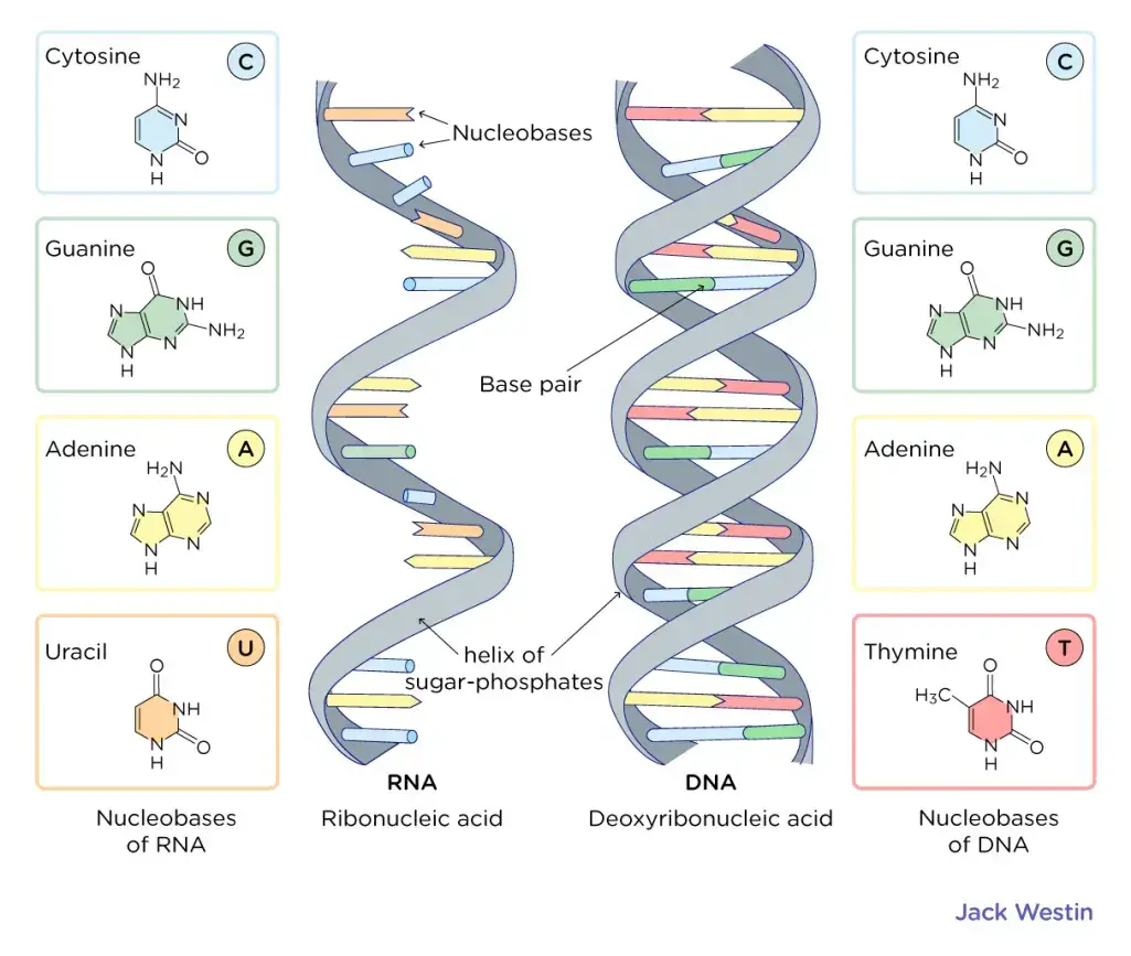
The Watson-Crick model of DNA, proposed by James Watson and Francis Crick, provides a comprehensive understanding of the structure and properties of DNA. Here is a summary of the key features of the Watson-Crick model:
- Double Helix: DNA is composed of two helical strands that coil around each other to form a double helix structure resembling a twisted ladder. The two strands are antiparallel, meaning they run in opposite directions.
- 5′ and 3′ Ends: Each DNA strand has a 5′ end and a 3′ end. The 5′ end of one strand is aligned with the 3′ end of the other strand.
- Diameter and Length: The DNA double helix has a uniform diameter of approximately 2 nanometers (nm). Each complete turn of the helix is about 3.6 nm in length, and there are 10.5 base pairs per turn.
- Base Pairing: The nitrogenous bases, Adenine (A), Thymine (T), Guanine (G), and Cytosine (C), are located inside the double helix. Adenine always pairs with Thymine through two hydrogen bonds, while Guanine always pairs with Cytosine through three hydrogen bonds.
- Major and Minor Grooves: The spiral arrangement of the two DNA strands creates distinct grooves. The major groove is larger and the minor groove is smaller. These grooves play a role in DNA-protein interactions and the binding of regulatory proteins.
- Complementary Base Pairs: The base pairing in DNA is highly specific, following the rule of complementary base pairs. A purine base (A or G) always pairs with a pyrimidine base (T or C), maintaining the balance between purines and pyrimidines.
- Hydrogen Bonds: Hydrogen bonds stabilize the base pairing between complementary bases in the double helix. Adenine-thymine pairs form two hydrogen bonds, while guanine-cytosine pairs form three hydrogen bonds.
- Sugar-Phosphate Backbone: The alternating sugar (deoxyribose) and phosphate groups form the backbone of the DNA double helix. The sugar-phosphate backbones are located on the exterior of the helix.
- Chargaff’s Rule: The base composition of DNA follows Chargaff’s rule, which states that the amount of Adenine is equal to Thymine, and the amount of Guanine is equal to Cytosine. This balance between the base pairs is crucial for the stability and proper functioning of DNA.
- Function and Replication: The structure of DNA elucidated by the Watson-Crick model has provided insights into DNA replication, gene expression, and protein synthesis. It serves as a template for DNA replication, transcription, and translation , enabling the transmission and utilization of genetic information in cells.
Overall, the Watson-Crick model of DNA has been instrumental in our understanding of DNA’s structure and function, paving the way for advances in genetics, molecular biology, and various fields of biological research.
DNA, or deoxyribonucleic acid, plays a fundamental role in biology and carries significant biological importance. Here are some key aspects of DNA’s biological significance:
- Genetic Information: DNA serves as the repository of genetic information in all living organisms. It contains the instructions that guide the development, growth, and functioning of an organism . The sequence of nucleotides in DNA determines the genetic code, which specifies the synthesis of proteins and governs the traits and characteristics of an organism.
- Inheritance : DNA is responsible for the inheritance of traits from parents to offspring. Through the process of reproduction, DNA is passed from one generation to the next, ensuring the transmission of genetic information. The unique combination of DNA sequences inherited from both parents contributes to the diversity and variability observed within and across species.
- Protein Synthesis: DNA serves as a template for protein synthesis. During the process of transcription, DNA is transcribed into messenger RNA ( mRNA ), which carries the genetic information to the ribosomes. Subsequently, during translation, the mRNA is used as a template to synthesize proteins, which perform essential functions in cells and organisms.
- Regulation of Gene Expression: DNA plays a crucial role in regulating gene expression, determining when and to what extent genes are turned on or off. Various mechanisms, such as DNA methylation and histone modification, can modify the structure and accessibility of DNA, influencing gene expression patterns. This regulation allows cells to respond to their environment and develop specialized functions.
- Evolution and Species Diversity: DNA underlies the process of evolution and the development of species diversity. Mutations, changes in the DNA sequence, introduce genetic variation , which can be acted upon by natural selection . Over time, these variations can lead to the emergence of new species and the adaptation of organisms to their environments.
- Forensic Identification: DNA analysis has become an invaluable tool in forensic science for identifying individuals. Each person’s DNA is unique, except for identical twins, making it a reliable method for establishing personal identity and determining relationships between individuals.
- Medical Applications: Understanding DNA has led to significant advancements in medicine. DNA sequencing and analysis help diagnose genetic disorders, predict disease susceptibility, and guide personalized treatments. DNA-based technologies, such as recombinant DNA and gene editing techniques like CRISPR-Cas9, hold promise for treating genetic diseases and developing new therapeutic approaches.
- Conservation and Biodiversity : DNA analysis is instrumental in conservation biology and the study of biodiversity. DNA barcoding allows for the identification and classification of species, including those that are endangered or difficult to distinguish based on morphology alone. DNA studies aid in understanding population dynamics, tracking migration patterns, and assessing the genetic health of species and ecosystems.
In summary, DNA’s biological importance lies in its role as the carrier of genetic information, its involvement in protein synthesis and gene regulation, its contribution to evolution and species diversity, its applications in forensics and medicine, and its significance in conservation and biodiversity studies. Understanding DNA has revolutionized our understanding of life and has far-reaching implications in various fields of science and technology.
The two strands of DNA are connected to each other through hydrogen bonds and a backbone of sugar-phosphate molecules.
The hydrogen bonds form between the nitrogenous bases of the two DNA strands. Adenine (A) always pairs with Thymine (T) through two hydrogen bonds, and Guanine (G) always pairs with Cytosine (C) through three hydrogen bonds. These specific base pairings ensure the complementary nature of the DNA strands.
The sugar-phosphate backbone runs along the outside of the double helix. It is composed of alternating deoxyribose sugar molecules and phosphate groups. The sugar molecules are linked together by phosphodiester bonds, which connect the 3′ carbon of one sugar to the 5′ carbon of the next sugar in the chain. This linkage creates a repeating sugar-phosphate backbone that provides stability and structure to the DNA molecule.
The combination of hydrogen bonds between the nitrogenous bases and the sugar-phosphate backbone holding the strands together forms the double helix structure of DNA. This connection allows for the precise pairing of the complementary bases on each strand and maintains the integrity of the genetic information encoded in the DNA molecule.
Chargaff’s rule, proposed by Erwin Chargaff, states that in a double-stranded DNA molecule, the amount of adenine (A) is equal to the amount of thymine (T), and the amount of guanine (G) is equal to the amount of cytosine (C). This rule can be summarized as A = T and G = C.
Chargaff’s rule is based on the observation that the composition of DNA varies between different species. By analyzing the base composition of DNA from different organisms, Chargaff discovered that the percentages of A and T are always approximately equal, as are the percentages of G and C. This finding was a crucial clue in understanding the structure and function of DNA.
The complementary base pairing in DNA, where A pairs with T and G pairs with C, is consistent with Chargaff’s rule. The pairing of these bases through hydrogen bonds ensures that the amount of purines (A and G) is always equal to the amount of pyrimidines (T and C), thus maintaining the structural integrity and stability of the DNA molecule.
Chargaff’s rule played a significant role in the discovery of the double helical structure of DNA by Watson and Crick. It provided valuable insight into the fundamental principles of DNA composition and helped establish the foundation for our understanding of the genetic code and DNA replication.
The key features of the Watson and Crick DNA model are as follows:
- Double Helix Structure : The model proposed by Watson and Crick describes DNA as a double-stranded helix. The two strands are coiled around each other in a twisted ladder-like structure.
- Antiparallel Strands: The two strands in the DNA molecule run in opposite directions. One strand has a 5′ end (with a phosphate group) and a 3′ end (with a hydroxyl group), while the other strand has a 3′ end and a 5′ end. This arrangement is known as antiparallel orientation.
- Complementary Base Pairing: The DNA strands are held together by complementary base pairing. Adenine (A) always pairs with thymine (T) through two hydrogen bonds, and guanine (G) always pairs with cytosine (C) through three hydrogen bonds. This pairing ensures the stability and integrity of the DNA molecule.
- Base Pairing Rule: T he base pairing rule in the Watson and Crick model states that the number of adenine (A) bases is equal to the number of thymine (T) bases, and the number of guanine (G) bases is equal to the number of cytosine (C) bases. This rule is also known as Chargaff’s rule.
- Sugar-Phosphate Backbone : The outside of the DNA double helix is formed by alternating sugar (deoxyribose) and phosphate groups. These sugar-phosphate backbones provide structural support and stability to the DNA molecule.
- Major and Minor Grooves: The twisting of the DNA double helix creates two grooves along its length. The major groove is wide and the minor groove is narrow. These grooves provide binding sites for proteins and other molecules involved in various cellular processes.
- Replication and Genetic Information: The Watson and Crick model of DNA provided insights into how DNA replicates and carries genetic information. The complementary base pairing allows for accurate replication, with each strand serving as a template for the synthesis of a new complementary strand.
- Molecular Basis of Inheritance: The discovery of the double helix structure of DNA by Watson and Crick laid the foundation for understanding the molecular basis of inheritance. It revealed how genetic information is encoded and transmitted in the form of DNA sequences.
These key features of the Watson and Crick DNA model have had a profound impact on our understanding of genetics, molecular biology, and the central role of DNA in life processes.
Chargaff’s rules played a crucial role in the development of the Watson and Crick model of DNA. Erwin Chargaff, a biochemist, discovered certain regularities in the composition of DNA bases, which provided important insights for understanding the structure of DNA. The contributions of Chargaff’s rules to the Watson and Crick model are as follows:
- Base Pairing: Chargaff’s rules stated that in DNA, the amount of adenine (A) is equal to the amount of thymine (T), and the amount of guanine (G) is equal to the amount of cytosine (C). This observation indicated a specific pairing between the bases, suggesting that A always pairs with T, and G always pairs with C.
- Complementary Base Pairing: Chargaff’s rules provided evidence for the concept of complementary base pairing in DNA. The equal ratios of A to T and G to C indicated that the bases are paired in a specific manner. This information was crucial for Watson and Crick in proposing their double helix model, where the complementary pairing of bases stabilizes the structure.
- Structural Constraints : Chargaff’s rules also implied that the total purine (A + G) content is equal to the total pyrimidine (T + C) content in DNA. This suggested that the DNA molecule had a consistent and uniform structure. Watson and Crick used this information to develop their model, ensuring that the double helix structure maintained equal spacing between the bases.
By following Chargaff’s rules, Watson and Crick were able to propose a model that incorporated the principles of complementary base pairing and the equal ratios of base pairs. Chargaff’s rules served as a guiding principle for understanding the structure and properties of DNA, and their contribution was instrumental in the development of the Watson and Crick model.
The discovery of the double helix structure by James Watson and Francis Crick in 1953 was a groundbreaking milestone in the field of molecular biology with immense significance. The key significance of their discovery can be summarized as follows:
- Understanding DNA Structure : Watson and Crick’s discovery provided the first accurate and comprehensive understanding of the three-dimensional structure of DNA. They proposed that DNA consists of two complementary strands that are twisted around each other in a double helix configuration. This knowledge laid the foundation for further research and exploration of the genetic material.
- Genetic Information Storage: The double helix structure revealed how genetic information is stored and transmitted in living organisms. The structure showed that the sequence of nucleotide bases along the DNA strands carries the genetic code, which determines the synthesis of proteins and other cellular functions. It established the connection between DNA structure and the hereditary characteristics of organisms.
- Base Pairing and Replication: Watson and Crick’s model demonstrated the specific base pairing of adenine (A) with thymine (T) and guanine (G) with cytosine (C). This base pairing provided insights into DNA replication, as it explained how each strand could serve as a template for the synthesis of a new complementary strand during cell division.
- Molecular Interactions: The discovery of the double helix structure highlighted the molecular interactions that stabilize the DNA molecule. It revealed that hydrogen bonds form between the base pairs, holding the two strands together. This understanding of molecular interactions in DNA paved the way for studying other biological macromolecules and their functions.
- Advances in Genetics and Biotechnology: The knowledge gained from the double helix structure has revolutionized the fields of genetics and biotechnology. It has enabled scientists to unravel the mechanisms of inheritance, gene expression, and genetic engineering . It has also facilitated advancements in DNA sequencing, genetic diagnostics, and the development of novel therapies and treatments.
- Impact on Scientific Community : Watson and Crick’s discovery sparked a scientific revolution and transformed our understanding of life at the molecular level. It inspired generations of scientists and paved the way for further discoveries in molecular biology, genetics, and related disciplines.
Overall, the significance of Watson and Crick’s discovery of the double helix structure lies in its profound impact on our understanding of DNA, genetics, and the fundamental mechanisms of life. It laid the groundwork for countless scientific breakthroughs and continues to shape research in biological sciences to this day.
The Watson and Crick model of DNA, also known as the double helix structure, provides a key explanation for the process of DNA replication. According to this model, DNA replication occurs through a semi-conservative mechanism, where each original DNA strand serves as a template for the synthesis of a new complementary strand. The process can be summarized as follows:
- Unwinding : The double-stranded DNA molecule unwinds and separates into two individual strands. This unwinding is facilitated by enzymes called DNA helicases, which break the hydrogen bonds between the base pairs and create a replication fork.
- Complementary Base Pairing: Each separated DNA strand serves as a template for the synthesis of a new complementary strand. The existing bases on the template strand determine the sequence of the new strand. Adenine (A) on the template strand pairs with thymine (T) and guanine (G) pairs with cytosine (C) through specific hydrogen bonding.
- DNA Polymerization: Enzymes known as DNA polymerases catalyze the addition of nucleotides to the growing DNA strand. DNA polymerase recognizes the exposed bases on the template strand and adds complementary nucleotides one by one, following the base-pairing rules. The nucleotides are joined together by phosphodiester bonds, forming the sugar-phosphate backbone of the new strand.
- Leading and Lagging Strands: DNA replication occurs in a continuous manner on one of the template strands, known as the leading strand. The DNA polymerase synthesizes the new complementary strand continuously in the 5′ to 3′ direction, following the replication fork. On the other template strand, called the lagging strand, replication occurs discontinuously in small fragments called Okazaki fragments. The fragments are later joined together by DNA ligase .
- Replication Fork Movement: As DNA replication progresses, the replication fork moves along the DNA molecule. The unwinding and synthesis of new strands occur simultaneously at the replication fork. Multiple replication forks can be present on a DNA molecule to expedite the replication process.
- Proofreading and Repair: DNA polymerases have proofreading capabilities to ensure the accuracy of replication. They can detect and correct errors in base pairing. Additionally, various repair mechanisms exist to fix any mistakes or damaged regions in the replicated DNA.
By following the Watson and Crick model, DNA replication ensures the faithful transmission of genetic information from one generation to the next. The model explains how each strand of the original DNA molecule serves as a template for the synthesis of new strands, resulting in two identical copies of the DNA molecule.
In the Watson and Crick DNA model, complementary base pairing plays a crucial role in the structure and replication of DNA. The model describes the double helix structure of DNA, where two strands of nucleotides are twisted around each other in a ladder-like arrangement.
Complementary base pairing refers to the specific pairing of nucleotide bases between the two DNA strands. In DNA, there are four nucleotide bases: adenine (A), thymine (T), cytosine (C), and guanine (G). The complementary base pairs are A with T and C with G. This means that adenine always pairs with thymine, and cytosine always pairs with guanine.
The base pairing is based on hydrogen bonding between the bases. Adenine forms two hydrogen bonds with thymine, while cytosine forms three hydrogen bonds with guanine. These hydrogen bonds provide stability to the DNA structure and hold the two strands together.
The complementary base pairing is significant because it allows for accurate DNA replication and the transmission of genetic information. During DNA replication, the two strands of the double helix separate, and each strand serves as a template for the synthesis of a new complementary strand. The base pairing rules ensure that the new strands are complementary to the original strands, resulting in two identical copies of the DNA molecule.
Additionally, the complementary base pairing plays a crucial role in gene expression and protein synthesis. The sequence of bases in DNA forms the genetic code, where each three-base sequence, called a codon, codes for a specific amino acid . The accurate base pairing ensures that the correct sequence of amino acids is encoded in the DNA and subsequently translated into proteins.
In summary, complementary base pairing in the Watson and Crick DNA model ensures the stability of the double helix structure, enables accurate DNA replication, and allows for the faithful transmission of genetic information.
Watson and Crick’s model of DNA was primarily based on a combination of existing experimental data and theoretical insights from other scientists. Although they did not conduct new experiments themselves, they incorporated crucial information from various sources to formulate their model. Some of the key evidence and data that contributed to their understanding of DNA are:
- X-ray crystallography by Rosalind Franklin and Maurice Wilkins: Franklin and Wilkins used X-ray crystallography techniques to study the structure of DNA fibers. Franklin’s X-ray diffraction images provided critical information about the helical nature of DNA and the spacing of its repeating units. Watson and Crick had access to Franklin’s unpublished data and utilized it to refine their model.
- Chargaff’s rules: Erwin Chargaff, a biochemist, discovered that in DNA, the amount of adenine (A) is equal to the amount of thymine (T), and the amount of cytosine (C) is equal to the amount of guanine (G). This observation, known as Chargaff’s rules, indicated a consistent pairing between the bases and provided hints about the structure of DNA.
- Linus Pauling’s alpha-helix structure: Linus Pauling, a prominent chemist, had proposed a helical structure for proteins based on their repeating patterns. Although Pauling’s alpha-helix structure was ultimately not applicable to DNA, it influenced Watson and Crick to consider a helical structure for DNA.
- Knowledge of chemical bonding: Watson and Crick had a strong background in chemistry, which helped them understand the principles of chemical bonding. They knew that hydrogen bonds were relatively weak but crucial for maintaining the stability of biological molecules. Applying this knowledge, they hypothesized that hydrogen bonding between specific base pairs could explain the complementarity of DNA strands.
By combining these pieces of information, Watson and Crick deduced the correct structure of DNA—a double helix with antiparallel strands held together by complementary base pairing. Their model elegantly explained how DNA could store and transmit genetic information, and it provided a solid foundation for understanding DNA replication and the central dogma of molecular biology.
The Watson and Crick model of DNA, proposed in 1953, revolutionized our understanding of genetics and molecular biology in several significant ways. Here are some key impacts of their model:
- Structure of DNA: Watson and Crick’s model revealed the double helix structure of DNA, which provided a physical framework for understanding how genetic information is stored and transmitted. It explained how the complementary base pairing allows for the accurate replication of DNA during cell division .
- DNA as the Genetic Material: The discovery of the DNA structure solidified the understanding that DNA is the molecule responsible for carrying genetic information. Prior to this, there were debates about whether proteins or nucleic acids were the carriers of genetic traits. The Watson and Crick model supported the notion that DNA, with its specific base sequences, encodes the instructions for protein synthesis and hereditary traits.
- Genetic Code and Protein Synthesis: The model’s base pairing rules and the understanding of the genetic code paved the way for deciphering how DNA sequences are translated into proteins. The codon-anticodon correspondence explained how specific combinations of three DNA bases (codons) specify the synthesis of particular amino acids during protein synthesis. This formed the basis for our understanding of gene expression and the central dogma of molecular biology.
- Heredity and Evolution: The Watson and Crick model provided insights into the mechanisms of heredity and evolution. It explained how DNA replication ensures the faithful transmission of genetic information from one generation to the next. It also shed light on the potential for genetic variations through mutations and recombination, which are the basis for genetic diversity and the driving forces behind evolution.
- Advances in Molecular Biology: The discovery of the DNA structure stimulated significant advancements in molecular biology. It opened doors for further research into DNA replication, repair, and recombination, as well as the regulation of gene expression. It also facilitated the development of techniques such as polymerase chain reaction ( PCR ), DNA sequencing, and genetic engineering, which have revolutionized various fields including medicine, agriculture, and biotechnology.
Overall, the Watson and Crick model of DNA provided a fundamental understanding of the molecular basis of genetics. It laid the groundwork for numerous breakthroughs in molecular biology, enabling us to unravel the mechanisms underlying inheritance, gene expression, and the intricate workings of living organisms.
- DNA can be used to create new materials. Scientists have been able to use DNA to create new materials with properties that are not found in naturally occurring materials. For example, DNA can be used to create materials that are transparent, conductive, and even self-healing.
- DNA can be used to create new drugs. Scientists are developing new drugs that use DNA as a delivery mechanism. These drugs can target specific cells or tissues , which could make them more effective and less harmful than traditional drugs.
- DNA can be used to create new sensors. Scientists are developing new sensors that use DNA to detect specific molecules. These sensors could be used to diagnose diseases, monitor environmental pollutants, and even track the movement of cells in the body.
- DNA can be used to create new biofuels. Scientists are developing new biofuels that are made from DNA. These biofuels could be used to replace fossil fuels, which would help to reduce greenhouse gas emissions.
- DNA can be used to create new batteries. Scientists are developing new batteries that are made from DNA. These batteries could be used to power devices that are too small for traditional batteries, such as implantable medical devices and wearable electronics.
- DNA can be used to create new electronics. Scientists are developing new electronics that are made from DNA. These electronics could be used to create new types of computers, sensors, and other devices.
- DNA can be used to create new DNA editing tools. Scientists are developing new DNA editing tools that use CRISPR-Cas9 technology. These tools could be used to correct genetic defects, create new crops and livestock, and even develop new forms of gene therapy .
- DNA can be used to create new vaccines . Scientists are developing new vaccines that use DNA as a delivery mechanism. These vaccines could be more effective and less expensive than traditional vaccines.
- DNA can be used to create new cancer treatments. Scientists are developing new cancer treatments that use DNA as a delivery mechanism. These treatments could target cancer cells more effectively and with fewer side effects than traditional treatments.
- DNA can be used to create new personalized medicine. Scientists are developing new personalized medicine approaches that use DNA to tailor treatments to individual patients. This could lead to more effective and less expensive treatments for a variety of diseases.
Watson and Crick discovered the double helix model of DNA.
Watson and Crick’s model of DNA showed that DNA is a double-stranded molecule made up of two helical chains spirally coiled around a common axis.
Watson and Crick’s DNA model can be described as a right-handed double helix structure with two antiparallel strands.
Drawing Watson and Crick’s model of DNA involves representing two strands coiled in a helical shape with complementary base pairing (adenine with thymine, and guanine with cytosine) and showing the sugar-phosphate backbones on the outside.
Chargaff’s rules helped Watson and Crick’s model of DNA by providing important insights into the base composition. Chargaff’s rule states that the amount of adenine is equal to thymine, and the amount of guanine is equal to cytosine, which guided Watson and Crick in understanding the complementary base pairing.
Watson and Crick developed their model of DNA through a combination of scientific data, analysis of X-ray crystallography images (such as those taken by Rosalind Franklin), and model building based on chemical principles.
Watson and Crick’s model of DNA featured a double helix structure with two strands, a uniform diameter of approximately 2 nanometers, and a repeating pattern of base pairs along the length.
Watson and Crick’s model of DNA demonstrated the complementary base pairing between adenine and thymine, and between guanine and cytosine, forming the steps of the DNA ladder.
Watson and Crick presented their DNA model on April 25, 1953, in a paper published in the scientific journal Nature titled “Molecular Structure of Nucleic Acids: A Structure for Deoxyribose Nucleic Acid.”
The first DNA model proposed by Watson and Crick had some inaccuracies and did not fully represent the precise structure of DNA. However, through continued research and collaboration, they refined their model to accurately depict the double helix structure.
- https://embryo.asu.edu/pages/genetical-implications-structure-deoxyribonucleic-acid-1953-james-watson-and-francis-crick
- https://www.nature.com/scitable/topicpage/discovery-of-dna-structure-and-function-watson-397/
- https://www.khanacademy.org/humanities/big-history-project/life/knowing-life/a/crick-watson-and-franklin2
- https://profiles.nlm.nih.gov/spotlight/sc/feature/doublehelix
- https://www.khanacademy.org/science/biology/dna-as-the-genetic-material/dna-discovery-and-structure/a/discovery-of-the-structure-of-dna
- https://thesciencenotes.com/watson-and-crick-double-helix-model-of-dna/
- https://unacademy.com/content/nta-ugc/study-material/pharmaceutical-analysis/the-watson-crick-structure-of-dna/
- https://ib.bioninja.com.au/standard-level/topic-2-molecular-biology/26-structure-of-dna-and-rna/watson–crick.html
- http://www.pbs.org/wgbh/aso/databank/entries/do53dn.html
- https://geneticeducation.co.in/dna-deoxyribonucleic-acid-definition-structure-function-evidence-and-types/
- https://www.mun.ca/biology/scarr/Watson-Crick_Model.html
- https://www.toppr.com/ask/question/explain-watson-and-cricks-model-of-dna/
- https://microbenotes.com/watson-and-crick-dna-model/
- https://byjus.com/neet/double-helix-structure-of-dna/
- https://www.merriam-webster.com/dictionary/Watson-Crick%20model
- https://www.vedantu.com/question-answer/explain-watson-and-crick-model-of-dna-class-12-biology-cbse-5f74594c0fd0025e7767ce48
- https://www.sciencehistory.org/historical-profile/james-watson-francis-crick-maurice-wilkins-and-rosalind-franklin
- https://study.com/academy/lesson/watson-crick-model-of-dna.html
- https://biologyreader.com/watson-and-crick-model-of-dna.html
- https://www.history.com/this-day-in-history/watson-and-crick-discover-chemical-structure-of-dna
- https://www.toppr.com/ask/en-us/question/explain-watson-and-cricks-model-of-dna/
- https://royalsociety.org/blog/2018/04/history-of-the-double-helix/
- https://easyhsc.com.au/home-easyhsc/easybio/heredity/cell-replication/watson-crick-dna-model/
- https://www.yourgenome.org/stories/unravelling-the-double-helix/
- http://www.lscollege.ac.in/sites/default/files/e-content/Structure%20of%20DNA.pdf
- https://jackwestin.com/resources/mcat-content/nucleic-acid-structure-and-function/deoxyribonucleic-acid-dna-double-helix-watson-crick-model-of-dna-structure
- https://www.javatpoint.com/dna-structure
- https://annex.exploratorium.edu/origins/coldspring/printit.html
- https://www.nobelprize.org/prizes/medicine/1962/speedread/
- https://dosequis.colorado.edu/Courses/MethodsLogic/papers/WatsonCrick1953.pdf
- https://undsci.berkeley.edu/the-structure-of-dna-cooperation-and-competition/a-false-start/
- https://www.zigya.com/study/book?class=11&board=cbse&subject=Biology&book=Biology&chapter=Biomolecules&q_type=&q_topic=Nature%20Of%20Bond%20Linking%20Monomers%20In%20A%20Polymer&question_id=BIEN11007981
- https://www.pbs.org/wgbh/evolution/library/06/3/l_063_01.html
- https://www.theguardian.com/science/2015/jun/23/sexism-in-science-did-watson-and-crick-really-steal-rosalind-franklins-data
- https://www.biologydiscussion.com/dna/watson-and-cricks-model-of-double-helix-of-dna-biochemistry/65076
- https://aklectures.com/lecture/structure-and-function/watson-crick-model-of-dna
- https://collection.sciencemuseumgroup.org.uk/objects/co146411/crick-and-watsons-dna-molecular-model-molecular-model
Related Biology Study Notes
Telomeres – structure, aging, shortening, functions, various model of replication – theta, rolling circle, and linear dna replication, theta model of replication – steps, applications, examples, what is dna replication – steps, enzymes, mechanism, applications, artificial selection – theory, types, advantages, examples, genetic variation – definition, types, causes, examples, gene expression and cell specialization – ap biology notes, eukaryotic gene regulation – mechanisms, regulatory elements, latest questions.
- All Questions
Start Asking Questions Cancel reply
Save my name, email, and website in this browser for the next time I comment.
This site uses Akismet to reduce spam. Learn how your comment data is processed .
- Click on your ad blocker icon in your browser's toolbar
- Select "Pause" or "Disable" for this website
- Refresh the page if it doesn't automatically reload

Understanding DNA: The Watson-Crick Model Explained
Explore the intricacies of DNA's structure and function through the Watson-Crick model, highlighting its role in genetic replication.

DNA is the cornerstone of genetic information, encoding the instructions vital for life. Its discovery and subsequent understanding have revolutionized fields such as genetics, medicine, and biotechnology. The Watson-Crick model, proposed in 1953, was a pivotal moment in molecular biology, providing clarity on DNA’s structural composition.
This article delves into the intricacies of the Watson-Crick model, shedding light on its components and significance.
Double Helix Structure
The double helix structure of DNA is a marvel of molecular architecture, characterized by its spiraling form. This configuration facilitates the storage and transmission of genetic information. The helical shape is formed by two long strands of nucleotides, which twist around each other like a ladder. Each strand is composed of a sugar-phosphate backbone, providing structural integrity and flexibility, allowing the helix to coil tightly within the confines of a cell nucleus.
The helical twist of DNA exhibits a specific pitch and diameter, which are important for its biological functions. The pitch, or the distance required for one complete turn of the helix, is approximately 3.4 nanometers, encompassing about ten nucleotide pairs. This measurement ensures that the genetic code is compactly organized, yet accessible for processes such as transcription and replication. The diameter of the helix, roughly 2 nanometers, is consistent, maintaining the stability of the structure under various cellular conditions.
Base Pairing Mechanism
Base pairing is at the heart of DNA’s ability to store and transmit genetic information with fidelity. This mechanism involves specific interactions between nitrogenous bases, which are the building blocks of the genetic code. These bases include adenine (A), thymine (T), guanine (G), and cytosine (C). The specificity of base pairing arises from the unique chemical structures of these bases, which allow them to form complementary pairs. Adenine pairs with thymine, while guanine pairs with cytosine. This complementary pairing ensures that each strand of the double helix can serve as a template for replication.
The pairing between these bases is stabilized by hydrogen bonds, which are weak interactions that, collectively, provide the strength required to hold the DNA strands together. Adenine and thymine are joined by two hydrogen bonds, whereas guanine and cytosine form three. This difference in bond number contributes to the overall stability of the DNA molecule, with G-C pairs being slightly more robust than A-T pairs. This stability is important during DNA replication and transcription, where strands must be separated to access the genetic code.
Antiparallel Strands
The concept of antiparallel strands is a defining feature of DNA’s structural elegance, contributing to its functional capabilities. Within the double helix, the two strands of DNA run in opposite directions, a configuration that is described as antiparallel. This orientation is dictated by the chemical directionality of the sugar-phosphate backbone, where one strand runs from the 5′ to the 3′ end, and the complementary strand runs from the 3′ to the 5′ end. This arrangement plays a significant role in the molecular interactions that underpin DNA’s biological functions.
The antiparallel nature of DNA strands is pivotal in the process of DNA replication. Enzymes responsible for replication, such as DNA polymerases, can only synthesize new DNA in a 5′ to 3′ direction. This directional requirement leads to the formation of the leading and lagging strands during replication. The leading strand is synthesized continuously, while the lagging strand is synthesized in short fragments known as Okazaki fragments, which are later joined together. This intricate process underscores the importance of the antiparallel orientation, ensuring that the genetic material is accurately copied.
Hydrogen Bonding
Hydrogen bonding plays a subtle yet indispensable role in the structural formation and stability of DNA. These bonds are the forces that maintain the fidelity of genetic information by holding the DNA strands together. Within the double helix, hydrogen bonds form between the nitrogenous bases, creating a robust yet flexible structure that allows the DNA to withstand various cellular processes. This flexibility permits the DNA to unwind and separate, enabling replication and transcription without compromising the integrity of the genetic code.
Hydrogen bonds confer specificity to base pairing, which is essential for accurate genetic replication and expression. The ability of hydrogen bonds to selectively form between specific bases ensures that the genetic code is consistently replicated without errors, maintaining the continuity of genetic information across generations. The relatively weak nature of individual hydrogen bonds allows for the dynamic processes of DNA repair and recombination, where temporary strand separation is required.
Major and Minor Grooves
The double helix structure of DNA contains distinct indentations known as major and minor grooves. These grooves are formed due to the asymmetric positioning of the sugar-phosphate backbones and the base pairs within the helix. The major groove is wider, providing more space for interactions with proteins and other molecules, while the minor groove is narrower. The presence of these grooves is critical for the binding of proteins that regulate gene expression and replication.
In the major groove, the larger surface area exposes more of the base pairs, allowing specific recognition by DNA-binding proteins such as transcription factors. These proteins can “read” the genetic information without unwinding the DNA, facilitating processes like gene activation and repression. The minor groove, though narrower, also plays a role in molecular recognition. Certain proteins and drugs can bind within this groove, influencing the structural conformation and biological activity of DNA. This dual-groove architecture enhances DNA’s ability to interact with a diverse array of molecules, contributing to the regulation and maintenance of genetic information.
Implications for Replication
The Watson-Crick model not only elucidates DNA’s structure but also provides insights into its replication mechanism. The antiparallel strands and base pairing mechanism ensure that each strand can serve as a template, allowing the genetic material to be accurately duplicated. During replication, the double helix unwinds, and replication forks are formed. Enzymes like helicase and DNA polymerase orchestrate the synthesis of new strands, ensuring that the genetic code is precisely transmitted to daughter cells. The semi-conservative nature of replication, where each new DNA molecule consists of one original and one new strand, underscores the reliability of this process.
DNA replication involves intricate regulation to prevent errors and ensure fidelity. Various checkpoints and repair mechanisms are in place to correct mismatches and damage. Enzymes such as exonucleases remove incorrect bases, while others like ligase seal the nicks in the phosphate backbone. This sophisticated system not only preserves genetic integrity but also allows for controlled variations, which are essential for evolution. The Watson-Crick model’s insights into replication continue to inform research in genetics and biotechnology, driving advances in areas such as genome editing and synthetic biology.

Key Features of the Genetic Code in Different Species
Genetic variability and cross-immunity in rsv strains, you may also be interested in..., genetic mechanisms and detection of penicillin resistance, chromosome dynamics and visualization in metaphase, microbial adaptation to environmental changes, advances in microbial genetics and cloning techniques.

Microbe Notes
Watson and Crick DNA Model
DNA stands for Deoxyribonucleic acid, a molecule that contains the instructions an organism needs to develop, live and reproduce. It is a type of nucleic acid and is one of the four major types of macromolecules that are known to be essential for all forms of life.
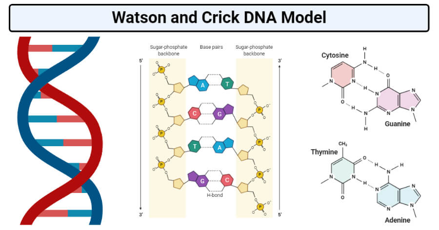
Table of Contents
Interesting Science Videos
- The three-dimensional structure of DNA, first proposed by James D. Watson and Francis H. C. Crick in 1953, consists of two long helical strands that are coiled around a common axis to form a double helix.
- Each DNA molecule is comprised of two biopolymer strands coiling around each other.
- Each strand has a 5′end (with a phosphate group) and a 3′end (with a hydroxyl group).
- The strands are antiparallel, meaning that one strand runs in a 5′to 3′direction, while the other strand runs in a 3′to 5′direction.
- The diameter of the double helix is 2nm and the double-helical structure repeats at an interval of 3.4nm which corresponds to ten base pairs.
- The two strands are held together by hydrogen bonds and are complementary to each other.
- The two DNA strands are called polynucleotides, as they are made of simpler monomer units called nucleotides. Basically, the DNA is composed of deoxyribonucleotides.
- The deoxyribonucleotides are linked together by 3′- 5′phosphodiester bonds.
- The nitrogenous bases that compose the deoxyribonucleotides include adenine, cytosine, thymine, and guanine.
- The structure of DNA -DNA is a double helix structure because it looks like a twisted ladder.
- The sides of the ladder are made of alternating sugar (deoxyribose) and phosphate molecules while the steps of the ladder are made up of a pair of nitrogen bases.
- As a result of the double-helical nature of DNA, the molecule has two asymmetric grooves. One groove is smaller than the other.
- This asymmetry is a result of the geometrical configuration of the bonds between the phosphate, sugar, and base groups that forces the base groups to attach at 120-degree angles instead of 180 degrees.
- The larger groove is called the major groove, occurs when the backbones are far apart; while the smaller one is called the minor groove, and occurs when they are close together.
- Since the major and minor grooves expose the edges of the bases, the grooves can be used to tell the base sequence of a specific DNA molecule.
- The possibility for such recognition is critical since proteins must be able to recognize specific DNA sequences on which to bind in order for the proper functions of the body and cell to be carried out.
Components of DNA Double Helix Structure
The Nitrogen Bases or Nucleotides
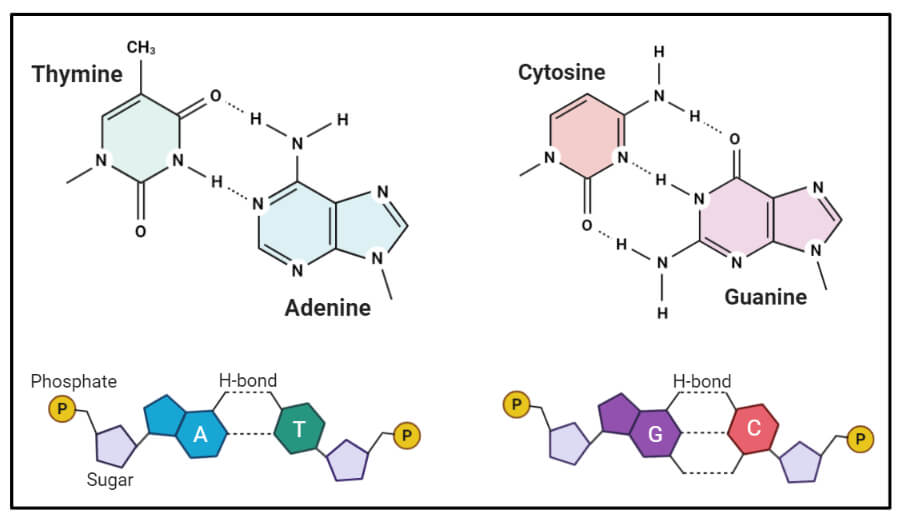
- DNA strands are composed of monomers called nucleotides.
- These monomers are often referred to as bases because they contain cyclic organic bases.
- Four different nucleotides, abbreviated A, T, C, and G, (adenine, thymine, cytosine, and guanine) are joined to form a DNA strand, with the base parts projecting inward from the backbone of the strand.
- Two strands bind together via the bases and twist to form a double helix.
- The nitrogen bases have a specific pairing pattern. This pairing pattern occurs because the amount of adenine equals the amount of thymine; the amount of guanine equals the amount of cytosine. The pairs are held together by hydrogen bonds.
- Each DNA double helix thus has a simple construction: wherever one strand has an A, the other strand has a T, and each C is matched with a G.
- The complementary strands are due to the nature of the nitrogenous bases. The base adenine always interacts with thymine (A-T) on the opposite strand via two hydrogen bonds and cytosine always interacts with guanine (C-G) via three hydrogen bonds on the opposite strand.
- The shape of the helix is stabilized by hydrogen bonding and hydrophobic interactions between bases.
Deoxyribose Sugar
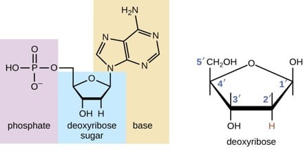
- Deoxyribose, also known as D-Deoxyribose and 2-deoxyribose, is a pentose sugar (monosaccharide containing five carbon atoms) that is a key component of the nucleic acid deoxyribonucleic acid (DNA).
- It is derived from the pentose sugar ribose. Deoxyribose has the chemical formula C 5 H 10 O 4 .
- Deoxyribose is the sugar component of DNA, just as ribose serves that role in RNA (ribonucleic acid).
- Alternating with phosphate bases, deoxyribose forms the backbone of the DNA, binding to the nitrogenous bases adenine, thymine, guanine, and cytosine.
- As a component of DNA, which represents the genetic information in all living cells, deoxyribose is critical to life. This ubiquitous sugar reflects a commonality among all living organisms.
The Phosphate Group (Phosphate Backbone)
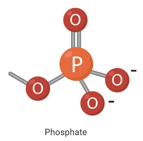
- The sugar-phosphate backbone forms the structural framework of nucleic acids, including DNA.
- This backbone is composed of alternating sugar and phosphate groups and defines the directionality of the molecule.
- DNA are composed of nucleotides that are linked to one another in a chain by chemical bonds, called ester bonds, between the sugar base of one nucleotide and the phosphate group of the adjacent nucleotide.
- The sugar is the 3′ end, and the phosphate is the 5′ end of each nucleotide.
- The phosphate group attached to the 5′ carbon of the sugar on one nucleotide forms an ester bond with the free hydroxyl on the 3′ carbon of the next nucleotide.
- These bonds are called phosphodiester bonds, and the sugar-phosphate backbone is described as extending, or growing, in the 5′ to 3′ direction when the molecule is synthesized.
- In double-stranded DNA, the molecular double-helix shape is formed by two linear sugar-phosphate backbones that run opposite each other and twist together in a helical shape.
- The sugar-phosphate backbone is negatively charged and hydrophilic, which allows the DNA backbone to form bonds with water.
- Alberts, B., Johnson, A., Lewis, J., Raff, M., Roberts, K., & Walter, P. (2002). Molecular biology of the cell. New York: Garland Science.
- http://www.newworldencyclopedia.org/entry/Deoxyribose
- https://www.nature.com/scitable/definition/phosphate-backbone-273
- https://www.slideshare.net/vinithaunnikrishnan16/forms-of-dna-49312507
- David Hames and Nigel Hooper (2005). Biochemistry. Third ed. Taylor & Francis Group: New York.
- Bailey, W. R., Scott, E. G., Finegold, S. M., & Baron, E. J. (1986). Bailey and Scott’s Diagnostic microbiology. St. Louis: Mosby.
About Author
Yashaswi Sharma
7 thoughts on “Watson and Crick DNA Model”
Surely, it was very helpful… thanks alot ????????
I am happy with your notes indeed
Surreal information. Explains the basic information to the best.
It was wonderful
It is very useful for me. Not only that i need it badly. But sir, if you tell me that if I have to write a short note about the watson and crick model of DNA, then what should I do? Are the first row of points are enough?
I like this place.
It’s really useful and easily understandable
Leave a Comment Cancel reply
Save my name, email, and website in this browser for the next time I comment.
This site uses Akismet to reduce spam. Learn how your comment data is processed .
Watson and Crick Model of DNA
Watson and Crick model of DNA provides one of the best ways to demonstrate the structure of double-helix DNA . A DNA is a polymer composed by the combination of several monomer units ( deoxyribonucleotides ) linked by the phosphodiester bond. In the discovery of DNA, many scientists have contextualized the structure of DNA, its components and composition etc.
Watson and Crick’s demonstrated a model, which explains all the physical and chemical features of the DNA. Before Watson and Crick, Friedrick Miescher, P. A. Levene, W.T. Astbury, Maurice Wilkins and Rosalind Franklin were the scientists who helped Watson and Crick to further study the DNA structure.
Watson and Crick’s model is the most successful model of DNA for which they have won the Noble Price in Physiology and Medicine in 1962 which they shared with Maurice Wilkins, but not with the Rosalind Franklin due to her unfortunate death in 1958.
Content: Watson and Crick Model of DNA
History of dna double helical structure, physical properties of dna, chemical properties of dna, in the year 1869.
A Swiss physiological chemist “ Freidrich Miescher ” identified something in the nuclei of the WBCs, which he termed it as “ Nuclein ”. Today, the term nuclein refers to a nucleic acid such as deoxyribonucleic acid.
In the year 1920
Phoebus Aaron Theodore Levene (an American organic chemist) discovered the elemental composition of nucleic acid. He explained one approach which is universally known as “ Tetranucleotide hypothesis ” according to which the base composition of all the four nitrogenous bases, i.e. A, T, G and C will be same.
In the year 1940
An English physicist and Molecular biologist, William Thomas Astbury gave the three dimensional model of DNA through X-ray crystallography.
In the year 1950
An Austro-Hungarian biochemist, Erwin Chargaff has demonstrated the “Biochemistry of DNA” by giving two approaches. Firstly, the sum of purines and the sum of pyrimidines will be equal in the DNA. Secondly, Erwin Chargaff concluded that the DNA composition varies between the different species. These two postulates commonly refer to the “ Chargaff’s Rule ”.
In the year 1952
Maurice Wilkins , Rosalind Franklin and co-workers introduced the photographs of the DNA by the method of “X-ray Diffraction”. R. Franklin has introduced the super X-ray diffraction photograph of DNA .
In the year 1953
Video: watson and crick model of dna.
Structure of DNA by Watson and Crick
Watson and Crick displayed the structure of DNA after studying the manuscript of the two scientists Linus Pauling and Corey. In 1953, Linus Pauling and Corey gave the 3D-structure of nucleic acid, which was not successful. Then, (in early 1953) Watson and Crick together combined the data of physical and chemical properties and proposed a double-helical structure of DNA. The main characteristics of Watson and Crick model of DNA include:
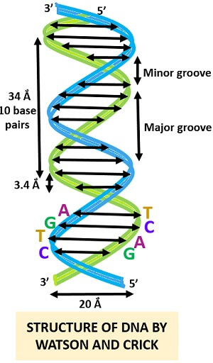
- According to the Watson and Crick model, the DNA is a double-stranded helix, which consists of two polynucleotide chains. The two polynucleotide chain are spirally or helically twisted, which gives it a twisted ladder-like look.
- Both the polynucleotide strands of DNA have the opposite polarities, which mean that the two strands will run in the antiparallel direction , i.e. one in 5’-3’ and other in 3’-5’ direction.
- The diameter of ds-stranded DNA helix is 20Å .
- The distance between the two nucleotides or internuclear distance is 3.4Å . The length of DNA helix is 34Å after a full turn and it possesses 10 base pairs per turn.
- The DNA is twisted in “Right-handed direction” or we can say in a “ Clockwise direction ”.
- Turning of DNA causes a formation of wide indentations, i.e. “ Major groove ”. The distance between the two strands forms a narrow indentation, i.e. “ Minor groove ”. The formation of major and minor grooves result after the DNA coiling and the grooves also act as a site of DNA binding proteins.
- There are four nucleotide bases present in the polynucleotide chain like adenine , guanine , cytosine and thymine . Adenine and guanine are the two purine bases, which have a single ring structure. Cytosine and thymine are the two pyrimidine bases, which have the double-ring structure.
- The two strands are joined together by the “ Complementary base pairing ” of the nitrogenous bases. Therefore, a purine base will complementarily pair with the pyrimidine base, in which ‘Adenine’ pairs with ‘Thymine’ and ‘Guanine’ pairs with ‘Cytosine’.
- The nucleotide bases in the polynucleotide strands of DNA will join with each other through a strong hydrogen bond .
- Adenine complementarily pairs with thymine through two hydrogen bonds , whereas guanine complementarily pairs with cytosine by means of three hydrogen bonds .
- The nucleotide base composition of DNA follows the Chargaff’s rule where the sum of purines is equal to the number of pyrimidines. The base composition of A + G = T + C obeys the Chargaff’s rule, but the base composition of A + T is not equal to the G + C.
- Polynucleotide strands of DNA consist of three major components, namely nitrogenous bases , deoxyribose sugar and a phosphate group.
- The backbone of DNA consists of the sugar-phosphate backbone. The sugar-phosphate backbone holds both the polynucleotide strands of DNA by means of “ Phosphodiester bond ”. Therefore, the bonding between sugar and phosphates, i.e. phosphodiester bond and the bonding between nitrogenous bases, i.e. hydrogen bond contributes to the “ DNA Stability ”.
The DNA is a supermodel proposed by Watson and Crick in the year 1953. The discovery of double helix DNA was not possible without the collaboration of Maurice Wilkins and Rosalind Franklin. Maurice Wilkins and Rosalind Franklin discovered the picture of DNA through X-ray crystallography . The X-ray diffraction picture of DNA helped Watson and Crick to further study the DNA structure and components. By this, Watson and Crick proposed a model for DNA known as Watson and Crick’s model of double-helical DNA .
The DNA is the largest biomolecule which contains all the genetic information of the person to build an organism or a life form. The study of DNA double-helical structure helps us to know about the chemical and physical properties of DNA, apart from the property of DNA being a “ Genetic material ”.
Related Topics:
- Growth Curve of Bacteria
- Precipitation Reaction
- Photoreactivation Repair
- Tobacco Mosaic Virus
- Difference Between Induction and Repression
2 thoughts on “Watson and Crick Model of DNA”
Thank you very much. It is so helpful for me.
Thank you in advance for the post. Its really made my assignment much easier.
Leave a Comment Cancel Reply
Your email address will not be published. Required fields are marked *
Start typing and press enter to search
“Molecular Structure of Nucleic Acids: A Structure for Deoxyribose Nucleic Acid” (1953), by James Watson and Francis Crick
In April 1953, James Watson and Francis Crick published "Molecular Structure of Nucleic Acids: A Structure of Deoxyribose Nucleic Acid" or "A Structure for Deoxyribose Nucleic Acid," in the journal Nature . In the article, Watson and Crick propose a novel structure for deoxyribonucleic acid or DNA. In 1944, Oswald T. Avery and his group at Rockefeller University in New York City, New York published experimental evidence that DNA contained the biological factors called genes that dictate how organisms grow and develop. Scientists did not know how DNAás function led to the passage of genetic information from cell to cell, or organism to organism. The model that Watson and Crick presented connected the concept of genes to heredity, growth, and development. As of 2018, most scientists accept Watson and Crick´s model of DNA presented in the article. For their work on DNA, Watson and Crick shared the 1962 Nobel Prize in Physiology or Medicine with Maurice Wilkins.
The collaboration that led Watson and Crick to write "A Structure of Deoxyribose Nucleic Acid" began in October 1951 soon after Watson arrived at the Cavendish Laboratory at the University of Cambridge in Cambridge, England. At the time, Watson was a twenty-three-year-old postdoctoral researcher from the United States and Crick, at the age of thirty-five, was a PhD student at the University of Cambridge. Watson and Crick started studying the structure of DNA together soon after Watson arrived at the Cavendish Laboratory. They frequently ate lunch together and discussed their work and the work of others in the laboratory. Eventually, senior members of the laboratory gave Watson and Crick an office space to share.
In 1944, Avery´s group at Rockefeller University Hospital published an article that provided experimental evidence that DNA contained genes. Decades before the publication of Watson and Crick´s article, scientists found evidence of the building blocks of DNA called nucleotides. Nucleotides are composed of three parts. The middle part of the nucleotide is a deoxyribose sugar attached to one side of the deoxyribose is a negatively charged phosphate group composed of phosphorus and oxygen and at the opposite side of the deoxyribose is one of four nitrogen bases, which varies between nucleotides. Scientists had published on the structure of the four bases in DNA: adenine, thymine, guanine, and cytosine. Adenine and guanine are made of two fused rings and called purines. Cytosine and thymine are single rings structures called pyrimidines.
When Watson and Crick wrote their article, they relied on the results of x-ray crystallography experiments. To conduct x-ray crystallography, scientists shoot a beam of x-rays, which are high-energy electromagnetic waves at a crystal. Once the beam hits the crystal, the x-rays scatter in a way that depends on the three-dimensional arrangements of the atoms in that crystal. The experiment results in an image, called a diffraction pattern, which scientists use to determine the three-dimensional structure of the crystal they observe. During the years leading up to the publication of the article, scientists used x-ray crystallography to learn about the three-dimensional structure of DNA. While Watson and Crick never conducted x-ray crystallography experiments themselves, they used data from experiments conducted by other scientists to develop their model of DNA.
Watson and Crick proposed a new model for the three-dimensional structure of DNA. The article consisted of less than two pages and had one illustration. To begin the paper, Watson and Crick respond to another DNA model proposed by Linus Pauling and Robert Corey, two scientists at the California Institute of Technology in Pasadena, California. Pauling and Corey proposed an alternate model just a few months before Watson and Crick wrote their article.
Watson and Crick begin their article by discussing the alternate model proposed by Pauling and Corey. While not described in detail by Watson and Crick, the Pauling-Corey model of DNA was a triple helix, where each of the three helical strands contained nucleotides strung together. In the Pauling-Corey model, the negatively charged phosphate groups faced inside the triple helix, and the bases faced outside the triple helix. In their article, Watson and Crick criticize the Pauling-Corey model. First, they argue that the DNA bases cannot face outward with the phosphate groups facing inward. Watson and Crick claim that the DNA strands bond together through the bases, so if the bases faced outward, there would be nothing connecting the DNA strands. The authors also argued that if the negatively charged phosphate backbones of the DNA strands faced inward they would repel each other. In addition, the arrangement of atoms in the Pauling-Corey structure would repel each other. That concludes Watson and Crick´s criticism of the Pauling-Corey model in their paper.
Watson and Crick continue with a description of their proposed DNA structure and a diagram to go along with their proposal. They define their structure as a double helix, with two helical chains coiled around a theoretical axis. According to Watson and Crick, the DNA strands run antiparallel to each other. That means that the strands run in opposite directions. Nucleotides are not perfectly symmetric molecules. They have a top and a bottom. So, when the DNA strands run antiparallel as Watson and Crick describe, one strand has the nucleotides facing right side up and the other strand has the nucleotides facing upside down.
Watson and Crick further the description of their structure by comparing it to a model of a chain of nucleic acids proposed by Sven Furberg, a crystallographer at Birbeck College, in London, United Kingdom, in 1952. Watson and Crick say that like Furberg´s model, their model of DNA has the bases facing inside the double helix and the phosphate backbones facing outside the double helix. Also similar to Furberg´s structure, Watson and Crick explain that the bases are perpendicular to the deoxyribose rings and phosphate groups of the nucleotides. In other words, the bases are like rungs of a ladder and the deoxyribose sugar and phosphate groups are like the rails of the ladder. After describing some remaining details about their structure, Watson and Crick finish their general description of their DNA model.
Watson and Crick then discuss the novel features pertaining to their DNA model. The first feature the authors discuss is how the two strands of DNA are connected. The authors state that a single base from one DNA strand attaches to a single base from the opposite DNA strand via hydrogen bonds. In DNA, hydrogen bonds occur between hydrogen atoms and oxygen or nitrogen atoms. While hydrogen bonds are weaker than the phosphate bonds connecting nucleotides together in each DNA strand, they are strong enough to hold the two helical strands together. Watson and Crick explain that for adequate hydrogen bonding to occur, within each pair of connected bases, one base must be a purine, a double ring, and one base must be a pyrimidine, a single ring.
Next, Watson and Crick describe is the specific identity of each base in a base pair. The authors assume that each of the four bases can only pair with one other type of base. Adenine, a purine, can only pair with thymine, a pyrimidine. Guanine, a purine, can only pair with cytosine, a pyrimidine. Based on that logic, Watson and Crick explain that the sequence of bases along one DNA strand automatically determines the sequence of the other strand. Each base along a DNA strand pairs with its only viable counterpart on the opposite strand. To support their claim about specific base paring, Watson and Crick cite experimental evidence. Erwin Chargaff at Columbia University in New York City, New York obtained that evidence. The authors explain that Chargaff determined that in DNA the ratio of adenine to thymine and guanine to cytosine is always roughly one-to-one. That means that the amount of adenine in DNA roughly equals the amount of thymine, and the amount of guanine roughly equals the amount of cytosine, which is the likely case if base pairing in DNA is specific.
After the discussion of base pairing, Watson and Crick conclude with x-ray crystallography evidence that they used to generate their model of DNA. Watson and Crick acknowledge that the x-ray crystallographic evidence of DNA published before they wrote their article could not confirm their model alone and there needs more experimental evidence to prove their model. Watson and Crick then postulate that the base pairing mechanism they proposed implied a possible DNA replication mechanism, though they do not describe that mechanism. The authors then end their article with acknowledgements.
When developing their model of DNA, Watson and Crick relied on unpublished x-ray crystallography experimental data. Scientists at King´s College London in London, UK, collected that data. Rosalind Franklin, a chemist, and her graduate student, Raymond Gosling, collected the data. Watson and Crick acknowledged those individuals in their paper. From 1951 to 1953, Franklin and Gosling gathered x-ray diffraction pattern images of DNA, which they obtained from the x-rays of DNA crystals. When Watson and Crick wrote "A Structure for Deoxyribose Nucleic Acid," Franklin and Gosling had not published their most clear DNA diffraction images, despite those images having improvements over the published data at the time. In early 1953, without Franklin´s knowledge, Maurice Wilkins a co-worker at King´s College showed Watson one of Franklin´s clear diffraction patterns of DNA. Later, Watson and Crick received a report Franklin wrote on her experimental findings. That report contained data Franklin presented at a colloquium at King´s College in 1951. When developing their own model of DNA, Watson and Crick drew conclusions from data contained within both Franklin´s diffraction image and her report.
In 1962, Watson and Crick shared the Nobel Prize in Physiology or Medicine with Wilkins for their findings relating to the structure of DNA and its role in genetics, many of which appeared in "A Structure for Deoxyribose Nucleic Acid." Franklin died in 1958 before the award of the 1962 Nobel Prize and did not receive the Nobel Prize, the award of the Nobel Prize is never posthumously. Some people speculate that if she were alive she would receive it for her contribution to solving the DNA structure. Others did not think she made crucial contributions to the solution of the DNA structure. The roll Franklin played in the discovery of the structure only realized after the publication of Watson´s book The Double Helix: A Personal Account of the Discovery of the Structure of DNA in 1968. Aaron Klug, Franklin´s last graduate student and colleague at Birkbeck College, in London, inherited her notebooks and papers when she died and published, "Rosalind Franklin and the Discovery of the Structure of DNA" a review of her notebooks and papers after publication of Watson´s book. Franklin and Gosling published five papers on DNA structure the first two sent to press before Franklin knew of the Watson-Crick Model. These papers were very technical papers dealing with x-ray crystallography, which may why they did not receive more attention.
Watson and Crick´s structure of DNA has remains largely accepted among scientists through the twenty-first century. The three-dimensional structure of DNA Watson and Crick proposed called the B-form DNA, terminology Franklin coined when she first collected diffraction patterns of that form of DNA. The B-form DNA is the most stable conformation of DNA under physiological conditions, though DNA can adopt other three-dimensional confirmations depending on its base sequence and its surrounding environment. For the next seven years following the 1953 publication of "A Structure of Deoxyribonucleic Acid," Wilkins and his research team obtained higher resolution x-ray diffraction images of B form DNA from a variety of species. From the higher quality images, Wilkins made small adjustments to the dimensions of Watson and Crick´s DNA structure.
"“A Structure of Deoxyribose Nucleic Acid" had immediate impacts on both the study of DNA as genetic material and the field of molecular biology. Watson and Crick´s article shifted scientists away from the question of how DNA was structured and toward the question of how DNA functioned. Later in 1953, Watson and Crick wrote a second paper, "Genetical Implications of the Structure of Deoxyribonucleic Acid," that addressed how DNA might self-replicate to pass on the genetic information encoded within it.
- Avery, Oswald T., Colin M. MacLeod, Maclyn McCarty. "Studies on the Chemical Nature of the Substance Inducing Transformation of Pneumococcal Types: Induction of Transformation by a Deoxyribonucleic Acid Fraction Isolated from Pneumococcus Type III" Journal of Experimental Medicine 79 (1944) 137–58. http://jem.rupress.org/content/79/2/137 (Accessed April 29, 2018).
- Chargaff, Erwin. "Chemical Specificity of Nucleic Acids and Mechanism of their Enzymatic Degradation." Cellular and Molecular Life Sciences 6 (1950): 201–9. http://biology.hunter.cuny.edu/molecularbio/Class%20Materials%20Spring%202012%20Biol302/Lecture%206/Chargaff.pdf (Accessed May 12, 2018).
- Furberg, Sven. "On the Structure of Nucleic Acids." Acta Chemica Scandinavia 6 (1952): 634–40. http://actachemscand.org/pdf/acta_vol_06_p0634-0640.pdf (Accessed May 12, 2018)
- Hamilton, Leonard D., Ralph K. Barclay, Maurice H. F. Wilkins, Geoffrey. L. Brown, Herbert R. Wilson, Donald A. Marvin, Harriett Ephrussi-Taylor, and Norman S. Simmons. "Similarity of the Structure of DNA from a Variety of Sources." The Journal of Cell Biology 5, (1959): 397–404. https://pdfs.semanticscholar.org/f547/465cb701c2e14573bd7c0ba62fb9c8aeb6c1.pdf (Accessed May 12, 2018).
- Judson, Horace Freeland. The Eighth Day of Creation . Cold Spring Harbor: Cold Spring Harbor Laboratory Press, 1996.
- Langridge, Robert, William E. Seeds, Herbert R. Wilson, Clive W. Hooper, Maurice H. F. Wilkins, and Leonard. D. Hamilton. "Molecular Structure of Deoxyribonucleic Acid (DNA)." The Journal of Biophysical and Biochemical Cytology 3 (1957): 767–78. https://www.ncbi.nlm.nih.gov/pmc/articles/PMC2224120/pdf/767.pdf . (Accessed May 12, 2018).
- Maddox, Brenda. Rosalind Franklin: The Dark Lady of DNA . London: HarperCollins Publishers, 2002.
- Maddox, Brenda. Rosalind Franklin: The Dark Lady of DNA. London: HarperCollins Publishers, 2002.
- Marsh, Richard E. "Biographical Memoir of Robert Brainard Corey". In Biographical Memoirs: National Academy of Sciences, Engineering, Medicine. Vol. 72 51–68. Washington D.C.: The National Academies Press, 1997.
- Maddox, Brenda. "The Double Helix and the ‘Wronged Heroine.’" Nature 421 (2003): 407–8. https://www.nature.com/articles/nature01399.pdf (Accessed May 4, 2018).
- Nelson, David L., Albert L. Lehninger, and Michael M. Cox. Lehninger Principles of Biochemistry. New York: Macmillan, 2008.
- Pauling, Linus, and Robert B. Corey. "A Proposed Structure for the Nucleic Acids." Proceedings of the National Academy of Sciences 39 (1953): 84–97. https://www.ncbi.nlm.nih.gov/pmc/articles/PMC1063734/ (Accessed May 21, 2018).
- Sayre, Anne. Rosalind Franklin and DNA. New York: W. W. Norton & Company, 1975.
- Watson, James D. The Double Helix: A Personal Account of the Discovery of the Structure of DNA. New York: Athenaeum Press, 1968. (Accessed May 12, 2018).
- Watson, James D., and Francis H.C. Crick. "Molecular Structure of Nucleic Acids." Nature 171 (1953): 737–8. https://www.genome.gov/edkit/pdfs/1953.pdf (Accessed May 12, 2018).
- Watson, James D., and Francis H.C. Crick. "Genetical Implications of the Structure of Deoxyribonucleic Acid." Nature 171 (1953): 964–7. https://www.nature.com/articles/171964b0 (Accessed April 29, 2018).
How to cite
Articles rights and graphics.
Copyright Arizona Board of Regents Licensed as Creative Commons Attribution-NonCommercial-Share Alike 3.0 Unported (CC BY-NC-SA 3.0)
Last modified
Share this page.

COMMENTS
Erwin Chargaff was one of a handful of scientists who expanded on Levene's work by uncovering additional details of the structure of DNA, thus further paving the way for Watson and Crick.
Watson and Crick published their findings in a one-page paper, with the understated title "A Structure for Deoxyribose Nucleic Acid," in the British scientific weekly Nature on April 25, 1953, illustrated with a schematic drawing of the double helix by Crick's wife, Odile. A coin toss decided the order in which they were named as authors.
May 31, 2024 · 1953: James Watson and Francis Crick, working in collaboration with Wilkins and Franklin, unveiled the double-helical structure of DNA. Watson and Crick’s model, based on the X-ray diffraction data, provided a groundbreaking understanding of how DNA is structured.
Oct 18, 2024 · The Watson-Crick model, proposed in 1953, was a pivotal moment in molecular biology, providing clarity on DNA’s structural composition. This article delves into the intricacies of the Watson-Crick model, shedding light on its components and significance. Double Helix Structure
Feb 1, 2022 · The three-dimensional structure of DNA, first proposed by James D. Watson and Francis H. C. Crick in 1953, consists of two long helical strands that are coiled around a common axis to form a double helix. Each DNA molecule is comprised of two biopolymer strands coiling around each other.
The X-ray diffraction picture of DNA helped Watson and Crick to further study the DNA structure and components. By this, Watson and Crick proposed a model for DNA known as Watson and Crick’s model of double-helical DNA. The DNA is the largest biomolecule which contains all the genetic information of the person to build an organism or a life form.
Watson and Crick took a crucial conceptual step, suggesting the molecule was made of two chains of nucleotides, each in a helix as Franklin had found, but one going up and the other going down.
May 1, 2023 · The Watson and Crick model of DNA, also known as the double helix model, is a scientific breakthrough that revolutionized our understanding of genetics. In 1953, James Watson and Francis Crick proposed that DNA has a double helix structure made up of two complementary strands, each consisting of a sugar-phosphate backbone and nitrogenous bases.
Francis Crick and James Watson with a model of the DNA molecule At midday on 28 February 1953, Francis Crick and James Watson walked into The Eagle pub in Cambridge and announced “We have ...
Oct 31, 2019 · While Watson and Crick never conducted x-ray crystallography experiments themselves, they used data from experiments conducted by other scientists to develop their model of DNA. Watson and Crick proposed a new model for the three-dimensional structure of DNA. The article consisted of less than two pages and had one illustration.